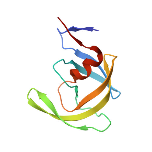
4U7Q

Structure of wild-type HIV protease in complex with photosensitive inhibitor PDI-6


Experimental Data Snapshot

  • Method: X-RAY DIFFRACTION
  • Resolution: 1.70 Å
  • R-Value Free: 0.326 
  • R-Value Work: 0.275 
  • R-Value Observed: 0.278 

wwPDB Validation   3D Report Full Report


Ligand Structure Quality Assessment 


This is version 2.1 of the entry. See complete history


Literature

Triggering HIV polyprotein processing by light using rapid photodegradation of a tight-binding protease inhibitor.

Schimer, J.Pavova, M.Anders, M.Pachl, P.Sacha, P.Cigler, P.Weber, J.Majer, P.Rezacova, P.Krausslich, H.G.Muller, B.Konvalinka, J.

(2015) Nat Commun 6: 6461-6461

  • DOI: https://doi.org/10.1038/ncomms7461
  • Primary Citation of Related Structures:  
    4U7Q, 4U7V

  • PubMed Abstract: 

    HIV protease (PR) is required for proteolytic maturation in the late phase of HIV replication and represents a prime therapeutic target. The regulation and kinetics of viral polyprotein processing and maturation are currently not understood in detail. Here we design, synthesize, validate and apply a potent, photodegradable HIV PR inhibitor to achieve synchronized induction of proteolysis. The compound exhibits subnanomolar inhibition in vitro. Its photolabile moiety is released on light irradiation, reducing the inhibitory potential by 4 orders of magnitude. We determine the structure of the PR-inhibitor complex, analyze its photolytic products, and show that the enzymatic activity of inhibited PR can be fully restored on inhibitor photolysis. We also demonstrate that proteolysis of immature HIV particles produced in the presence of the inhibitor can be rapidly triggered by light enabling thus to analyze the timing, regulation and spatial requirements of viral processing in real time.


  • Organizational Affiliation

    1] Institute of Organic Chemistry and Biochemistry, Academy of Sciences of the Czech Republic, Gilead Sciences and IOCB Research Center, Flemingovo n.2, 166 10, Prague 6, Czech Republic [2] Department of Biochemistry, Faculty of Science, Charles University in Prague, Hlavova 8, 128 43, Prague 2, Czech Republic.


Macromolecules
Find similar proteins by:  (by identity cutoff)  |  3D Structure
Entity ID: 1
MoleculeChains Sequence LengthOrganismDetailsImage
V-1 protease
A, B
99Human immunodeficiency virus 1Mutation(s): 0 
Gene Names: ORF
UniProt
Find proteins for P04585 (Human immunodeficiency virus type 1 group M subtype B (isolate HXB2))
Explore P04585 
Go to UniProtKB:  P04585
Entity Groups  
Sequence Clusters30% Identity50% Identity70% Identity90% Identity95% Identity100% Identity
UniProt GroupP04585
Sequence Annotations
Expand
  • Reference Sequence
Small Molecules
Ligands 1 Unique
IDChains Name / Formula / InChI Key2D Diagram3D Interactions
3EM
Query on 3EM

Download Ideal Coordinates CCD File 
C [auth B]N~2~-({[7-(diethylamino)-2-oxo-2H-chromen-4-yl]methoxy}carbonyl)-N-[(2S,4S,5S)-4-hydroxy-1,6-diphenyl-5-{[(1,3-thiazol-5-ylmethoxy)carbonyl]amino}hexan-2-yl]-L-valinamide
C43 H51 N5 O8 S
HWKZGVMRBIGKNW-FUVWEXRVSA-N
Binding Affinity Annotations 
IDSourceBinding Affinity
3EM Binding MOAD:  4U7Q Ki: 0.18 (nM) from 1 assay(s)
Experimental Data & Validation

Experimental Data

  • Method: X-RAY DIFFRACTION
  • Resolution: 1.70 Å
  • R-Value Free: 0.326 
  • R-Value Work: 0.275 
  • R-Value Observed: 0.278 
  • Space Group: P 61
Unit Cell:
Length ( Å )Angle ( ˚ )
a = 62.841α = 90
b = 62.841β = 90
c = 80.486γ = 120
Software Package:
Software NamePurpose
XDSdata reduction
PDB_EXTRACTdata extraction
MOLREPphasing
REFMACrefinement
XSCALEdata scaling

Structure Validation

View Full Validation Report


Ligand Structure Quality Assessment 


Entry History & Funding Information

Deposition Data


Funding OrganizationLocationGrant Number
Ministry of Education of the CzechRepublicCzech RepublicP208-12-G016
Ministry of Education, Youth and Sports of the Czech RepublicCzech RepublicLO 1302
German Research FoundationGermanyMU885/5-1

Revision History  (Full details and data files)

  • Version 1.0: 2015-03-25
    Type: Initial release
  • Version 2.0: 2017-09-06
    Changes: Atomic model, Author supporting evidence, Derived calculations, Refinement description
  • Version 2.1: 2023-12-20
    Changes: Data collection, Database references, Refinement description