4TVH

HIV Protease (PR) dimer in closed form with TL-3 in active site and fragment AK-2097 in the outside/top of flap


Experimental Data Snapshot

  • Method: X-RAY DIFFRACTION
  • Resolution: 2.19 Å
  • R-Value Free: 0.260 
  • R-Value Work: 0.184 
  • R-Value Observed: 0.187 

wwPDB Validation   3D Report Full Report


Ligand Structure Quality Assessment 


This is version 1.5 of the entry. See complete history


Literature

Distinguishing Binders from False Positives by Free Energy Calculations: Fragment Screening Against the Flap Site of HIV Protease.

Deng, N.Forli, S.He, P.Perryman, A.Wickstrom, L.Vijayan, R.S.Tiefenbrunn, T.Stout, D.Gallicchio, E.Olson, A.J.Levy, R.M.

(2015) J Phys Chem B 119: 976-988

  • DOI: https://doi.org/10.1021/jp506376z
  • Primary Citation of Related Structures:  
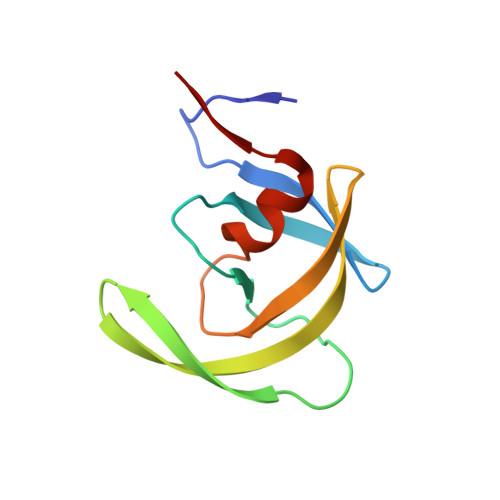
    4TVG, 4TVH

  • PubMed Abstract: 

    Molecular docking is a powerful tool used in drug discovery and structural biology for predicting the structures of ligand-receptor complexes. However, the accuracy of docking calculations can be limited by factors such as the neglect of protein reorganization in the scoring function; as a result, ligand screening can produce a high rate of false positive hits. Although absolute binding free energy methods still have difficulty in accurately rank-ordering binders, we believe that they can be fruitfully employed to distinguish binders from nonbinders and reduce the false positive rate. Here we study a set of ligands that dock favorably to a newly discovered, potentially allosteric site on the flap of HIV-1 protease. Fragment binding to this site stabilizes a closed form of protease, which could be exploited for the design of allosteric inhibitors. Twenty-three top-ranked protein-ligand complexes from AutoDock were subject to the free energy screening using two methods, the recently developed binding energy analysis method (BEDAM) and the standard double decoupling method (DDM). Free energy calculations correctly identified most of the false positives (≥83%) and recovered all the confirmed binders. The results show a gap averaging ≥3.7 kcal/mol, separating the binders and the false positives. We present a formula that decomposes the binding free energy into contributions from the receptor conformational macrostates, which provides insights into the roles of different binding modes. Our binding free energy component analysis further suggests that improving the treatment for the desolvation penalty associated with the unfulfilled polar groups could reduce the rate of false positive hits in docking. The current study demonstrates that the combination of docking with free energy methods can be very useful for more accurate ligand screening against valuable drug targets.


  • Organizational Affiliation

    Center for Biophysics & Computational Biology/ICMS, ‡Department of Chemistry, Temple University , Philadelphia, Pennsylvania19122, United States.


Macromolecules
Find similar proteins by:  (by identity cutoff)  |  3D Structure
Entity ID: 1
MoleculeChains Sequence LengthOrganismDetailsImage
Protease
A, B
99Human immunodeficiency virus 1Mutation(s): 1 
Gene Names: pol
UniProt
Find proteins for Q90EA1 (Human immunodeficiency virus 1)
Explore Q90EA1 
Go to UniProtKB:  Q90EA1
Entity Groups  
Sequence Clusters30% Identity50% Identity70% Identity90% Identity95% Identity100% Identity
UniProt GroupQ90EA1
Sequence Annotations
Expand
  • Reference Sequence
Small Molecules
Ligands 3 Unique
IDChains Name / Formula / InChI Key2D Diagram3D Interactions
3TL
Query on 3TL

Download Ideal Coordinates CCD File 
C [auth A]benzyl [(1S,4S,7S,8R,9R,10S,13S,16S)-7,10-dibenzyl-8,9-dihydroxy-1,16-dimethyl-4,13-bis(1-methylethyl)-2,5,12,15,18-pentaoxo-20-phenyl-19-oxa-3,6,11,14,17-pentaazaicos-1-yl]carbamate
C50 H64 N6 O10
BJJPNOGMLLUCER-KUTQPOQPSA-N
K97
Query on K97

Download Ideal Coordinates CCD File 
E [auth B]3-(morpholin-4-ylmethyl)-1H-indole-6-carboxylic acid
C14 H16 N2 O3
PGNDNJYNINUQIJ-UHFFFAOYSA-N
BME
Query on BME

Download Ideal Coordinates CCD File 
D [auth B]BETA-MERCAPTOETHANOL
C2 H6 O S
DGVVWUTYPXICAM-UHFFFAOYSA-N
Binding Affinity Annotations 
IDSourceBinding Affinity
3TL BindingDB:  4TVH Ki: 1.5 (nM) from 1 assay(s)
Biologically Interesting Molecules (External Reference) 1 Unique
Experimental Data & Validation

Experimental Data

  • Method: X-RAY DIFFRACTION
  • Resolution: 2.19 Å
  • R-Value Free: 0.260 
  • R-Value Work: 0.184 
  • R-Value Observed: 0.187 
  • Space Group: P 21 21 21
Unit Cell:
Length ( Å )Angle ( ˚ )
a = 28.916α = 90
b = 66.295β = 90
c = 92.175γ = 90
Software Package:
Software NamePurpose
SCALAdata scaling
REFMACrefinement
PDB_EXTRACTdata extraction
PHASERphasing
PROTEUMdata reduction

Structure Validation

View Full Validation Report


Ligand Structure Quality Assessment 


Entry History & Funding Information

Deposition Data


Funding OrganizationLocationGrant Number
National Institutes of Health/National Institute of General Medical Sciences (NIH/NIGMS)United StatesGM103368-02

Revision History  (Full details and data files)

  • Version 1.0: 2014-09-17
    Type: Initial release
  • Version 1.1: 2015-02-04
    Changes: Database references
  • Version 1.2: 2017-09-13
    Changes: Author supporting evidence, Database references, Derived calculations, Other, Refinement description, Source and taxonomy
  • Version 1.3: 2017-11-22
    Changes: Refinement description
  • Version 1.4: 2019-12-25
    Changes: Author supporting evidence
  • Version 1.5: 2023-09-27
    Changes: Data collection, Database references, Refinement description