4TR2

Crystal structure of PvSUB1


Experimental Data Snapshot

  • Method: X-RAY DIFFRACTION
  • Resolution: 2.70 Å
  • R-Value Free: 0.244 
  • R-Value Work: 0.200 
  • R-Value Observed: 0.202 

wwPDB Validation   3D Report Full Report


This is version 2.1 of the entry. See complete history


Literature

A novel Plasmodium-specific prodomain fold regulates the malaria drug target SUB1 subtilase.

Giganti, D.Bouillon, A.Tawk, L.Robert, F.Martinez, M.Crublet, E.Weber, P.Girard-Blanc, C.Petres, S.Haouz, A.Hernandez, J.F.Mercereau-Puijalon, O.Alzari, P.M.Barale, J.C.

(2014) Nat Commun 5: 4833-4833

  • DOI: https://doi.org/10.1038/ncomms5833
  • Primary Citation of Related Structures:  
    4TR2

  • PubMed Abstract: 

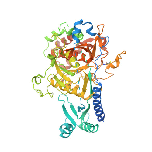
    The Plasmodium subtilase SUB1 plays a pivotal role during the egress of malaria parasites from host hepatocytes and erythrocytes. Here we report the crystal structure of full-length SUB1 from the human-infecting parasite Plasmodium vivax, revealing a bacterial-like catalytic domain in complex with a Plasmodium-specific prodomain. The latter displays a novel architecture with an amino-terminal insertion that functions as a 'belt', embracing the catalytic domain to further stabilize the quaternary structure of the pre-protease, and undergoes calcium-dependent autoprocessing during subsequent activation. Although dispensable for recombinant enzymatic activity, the SUB1 'belt' could not be deleted in Plasmodium berghei, suggesting an essential role of this domain for parasite development in vivo. The SUB1 structure not only provides a valuable platform to develop new anti-malarial candidates against this promising drug target, but also defines the Plasmodium-specific 'belt' domain as a key calcium-dependent regulator of SUB1 during parasite egress from host cells.


  • Organizational Affiliation

    1] Institut Pasteur, Unité de Microbiologie Structurale, Département de Biologie Structurale et Chimie, F-75015 Paris, France [2] CNRS UMR 3528, F-75015 Paris, France.


Macromolecules
Find similar proteins by:  (by identity cutoff)  |  3D Structure
Entity ID: 1
MoleculeChains Sequence LengthOrganismDetailsImage
Subtilisin-like 1 serine protease
A, B
663Plasmodium vivaxMutation(s): 0 
Gene Names: sub1
UniProt
Find proteins for E6Y8B9 (Plasmodium vivax)
Explore E6Y8B9 
Go to UniProtKB:  E6Y8B9
Entity Groups  
Sequence Clusters30% Identity50% Identity70% Identity90% Identity95% Identity100% Identity
UniProt GroupE6Y8B9
Sequence Annotations
Expand
  • Reference Sequence
Small Molecules
Ligands 2 Unique
IDChains Name / Formula / InChI Key2D Diagram3D Interactions
PO4
Query on PO4

Download Ideal Coordinates CCD File 
G [auth A],
H [auth A],
I [auth A],
N [auth B],
O [auth B]
PHOSPHATE ION
O4 P
NBIIXXVUZAFLBC-UHFFFAOYSA-K
CA
Query on CA

Download Ideal Coordinates CCD File 
C [auth A]
D [auth A]
E [auth A]
F [auth A]
J [auth B]
C [auth A],
D [auth A],
E [auth A],
F [auth A],
J [auth B],
K [auth B],
L [auth B],
M [auth B]
CALCIUM ION
Ca
BHPQYMZQTOCNFJ-UHFFFAOYSA-N
Experimental Data & Validation

Experimental Data

  • Method: X-RAY DIFFRACTION
  • Resolution: 2.70 Å
  • R-Value Free: 0.244 
  • R-Value Work: 0.200 
  • R-Value Observed: 0.202 
  • Space Group: P 43 21 2
Unit Cell:
Length ( Å )Angle ( ˚ )
a = 95α = 90
b = 95β = 90
c = 287.76γ = 90
Software Package:
Software NamePurpose
BUSTERrefinement

Structure Validation

View Full Validation Report


Entry History & Funding Information

Deposition Data


Funding OrganizationLocationGrant Number
French National Research AgencyFranceANR-11-RPIB-002

Revision History  (Full details and data files)

  • Version 1.0: 2014-09-17
    Type: Initial release
  • Version 1.1: 2014-09-24
    Changes: Database references
  • Version 2.0: 2017-08-30
    Changes: Atomic model, Author supporting evidence, Derived calculations
  • Version 2.1: 2023-12-20
    Changes: Data collection, Database references, Derived calculations, Refinement description