4THI

THIAMINASE I FROM BACILLUS THIAMINOLYTICUS WITH COVALENTLY BOUND 4-AMINO-2,5-DIMETHYLPYRIMIDINE


Experimental Data Snapshot

  • Method: X-RAY DIFFRACTION
  • Resolution: 2.00 Å
  • R-Value Free: 0.258 
  • R-Value Work: 0.211 
  • R-Value Observed: 0.211 

wwPDB Validation   3D Report Full Report


This is version 1.3 of the entry. See complete history


Literature

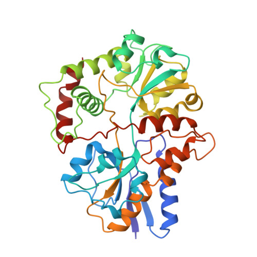
Crystal structure of thiaminase-I from Bacillus thiaminolyticus at 2.0 A resolution.

Campobasso, N.Costello, C.A.Kinsland, C.Begley, T.P.Ealick, S.E.

(1998) Biochemistry 37: 15981-15989

  • DOI: https://doi.org/10.1021/bi981673l
  • Primary Citation of Related Structures:  
    2THI, 3THI, 4THI

  • PubMed Abstract: 

    Thiaminase-I catalyzes the replacement of the thiazole moiety of thiamin with a wide variety of nucleophiles, such as pyridine, aniline, catechols, quinoline, and cysteine. The crystal structure of the enzyme from Bacillus thiaminolyticus was determined at 2.5 A resolution by multiple isomorphous replacement and refined to an R factor of 0.195 (Rfree = 0.272). Two other structures, one native and one containing a covalently bound inhibitor, were determined at 2.0 A resolution by molecular replacement from a second crystal form and were refined to R factors of 0.205 and 0.217 (Rfree = 0.255 and 0.263), respectively. The overall structure contains two alpha/beta-type domains separated by a large cleft. At the base of the cleft lies Cys113, previously identified as a key active site nucleophile. The structure with a covalently bound thiamin analogue, which functions as a mechanism-based inactivating agent, confirms the location of the active site. Glu241 appears to function as an active site base to increase the nucleophilicity of Cys113. The mutant Glu241Gln was made and shows no activity. Thiaminase-I shows no sequence identity to other proteins in the sequence databases, but the three-dimensional structure shows very high structural homology to the periplasmic binding proteins and the transferrins.


  • Organizational Affiliation

    Department of Chemistry and Chemical Biology, Cornell University, Ithaca, New York 14853, USA.


Macromolecules
Find similar proteins by:  (by identity cutoff)  |  3D Structure
Entity ID: 1
MoleculeChains Sequence LengthOrganismDetailsImage
PROTEIN (THIAMINASE I)362Bacillus subtilisMutation(s): 0 
EC: 2.5.1.2
UniProt
Find proteins for P45741 (Paenibacillus thiaminolyticus)
Explore P45741 
Go to UniProtKB:  P45741
Entity Groups  
Sequence Clusters30% Identity50% Identity70% Identity90% Identity95% Identity100% Identity
UniProt GroupP45741
Sequence Annotations
Expand
  • Reference Sequence
Small Molecules
Ligands 2 Unique
IDChains Name / Formula / InChI Key2D Diagram3D Interactions
PYD
Query on PYD

Download Ideal Coordinates CCD File 
C [auth A]2,5-DIMETHYL-PYRIMIDIN-4-YLAMINE
C6 H9 N3
UXKNAXNFIYFMIB-UHFFFAOYSA-N
SO4
Query on SO4

Download Ideal Coordinates CCD File 
B [auth A]SULFATE ION
O4 S
QAOWNCQODCNURD-UHFFFAOYSA-L
Experimental Data & Validation

Experimental Data

  • Method: X-RAY DIFFRACTION
  • Resolution: 2.00 Å
  • R-Value Free: 0.258 
  • R-Value Work: 0.211 
  • R-Value Observed: 0.211 
  • Space Group: P 21 21 2
Unit Cell:
Length ( Å )Angle ( ˚ )
a = 86.7α = 90
b = 119.3β = 90
c = 37.3γ = 90
Software Package:
Software NamePurpose
X-PLORmodel building
X-PLORrefinement
DENZOdata reduction
SCALEPACKdata scaling
X-PLORphasing

Structure Validation

View Full Validation Report


Entry History 

Deposition Data

Revision History  (Full details and data files)

  • Version 1.0: 1998-10-14
    Type: Initial release
  • Version 1.1: 2007-10-16
    Changes: Version format compliance
  • Version 1.2: 2011-07-13
    Changes: Version format compliance
  • Version 1.3: 2023-09-20
    Changes: Data collection, Database references, Derived calculations, Refinement description