4S2S

Crystal Structure of Fab fragment of monoclonal antibody RoAb13


Experimental Data Snapshot

  • Method: X-RAY DIFFRACTION
  • Resolution: 2.10 Å
  • R-Value Free: 0.234 
  • R-Value Work: 0.186 
  • R-Value Observed: 0.189 

wwPDB Validation   3D Report Full Report


This is version 1.1 of the entry. See complete history


Literature

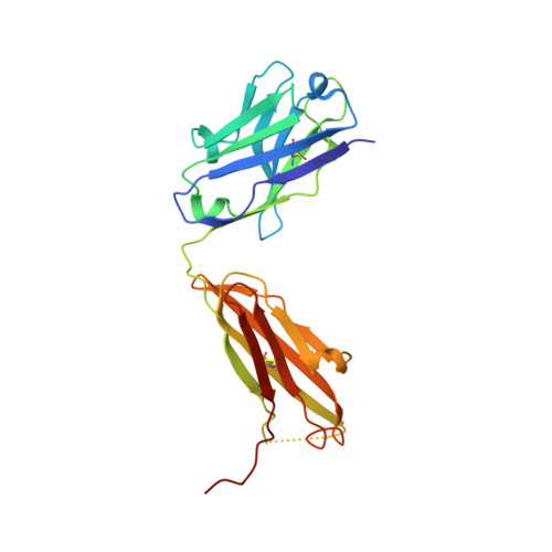
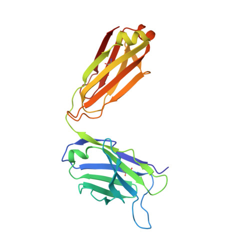
A Linear Epitope in the N-Terminal Domain of CCR5 and Its Interaction with Antibody.

Chain, B.Arnold, J.Akthar, S.Brandt, M.Davis, D.Noursadeghi, M.Lapp, T.Ji, C.Sankuratri, S.Zhang, Y.Govada, L.Saridakis, E.Chayen, N.

(2015) PLoS One 10: e0128381-e0128381

  • DOI: https://doi.org/10.1371/journal.pone.0128381
  • Primary Citation of Related Structures:  
    4S2S

  • PubMed Abstract: 

    The CCR5 receptor plays a role in several key physiological and pathological processes and is an important therapeutic target. Inhibition of the CCR5 axis by passive or active immunisation offers one very selective strategy for intervention. In this study we define a new linear epitope within the extracellular domain of CCR5 recognised by two independently produced monoclonal antibodies. A short peptide encoding the linear epitope can induce antibodies which recognise the intact receptor when administered colinear with a tetanus toxoid helper T cell epitope. The monoclonal antibody RoAb 13 is shown to bind to both cells and peptide with moderate to high affinity (6x10^8 and 1.2x10⁷ M-1 respectively), and binding to the peptide is enhanced by sulfation of tyrosines at positions 10 and 14. RoAb13, which has previously been shown to block HIV infection, also blocks migration of monocytes in response to CCR5 binding chemokines and to inflammatory macrophage conditioned medium. A Fab fragment of RoAb13 has been crystallised and a structure of the antibody is reported to 2.1 angstrom resolution.


  • Organizational Affiliation

    Division of Infection and Immunity, UCL, Gower St., London, United Kingdom.


Macromolecules
Find similar proteins by:  (by identity cutoff)  |  3D Structure
Entity ID: 1
MoleculeChains Sequence LengthOrganismDetailsImage
RoAb13 Fab Heavy chainA [auth H],
C [auth A]
224Mus musculusMutation(s): 0 
Entity Groups  
Sequence Clusters30% Identity50% Identity70% Identity90% Identity95% Identity100% Identity
Sequence Annotations
Expand
  • Reference Sequence
Find similar proteins by:  (by identity cutoff)  |  3D Structure
Entity ID: 2
MoleculeChains Sequence LengthOrganismDetailsImage
RoAb13 Fab Light chainB [auth L],
D [auth B]
217Mus musculusMutation(s): 0 
Entity Groups  
Sequence Clusters30% Identity50% Identity70% Identity90% Identity95% Identity100% Identity
Sequence Annotations
Expand
  • Reference Sequence
Experimental Data & Validation

Experimental Data

  • Method: X-RAY DIFFRACTION
  • Resolution: 2.10 Å
  • R-Value Free: 0.234 
  • R-Value Work: 0.186 
  • R-Value Observed: 0.189 
  • Space Group: P 1 21 1
Unit Cell:
Length ( Å )Angle ( ˚ )
a = 75.2α = 90
b = 88.64β = 103.19
c = 78.45γ = 90
Software Package:
Software NamePurpose
CrystalCleardata collection
MOLREPphasing
PHENIXrefinement
MOSFLMdata reduction
SCALAdata scaling

Structure Validation

View Full Validation Report


Entry History 

Deposition Data

Revision History  (Full details and data files)

  • Version 1.0: 2015-06-24
    Type: Initial release
  • Version 1.1: 2023-09-20
    Changes: Data collection, Database references, Refinement description