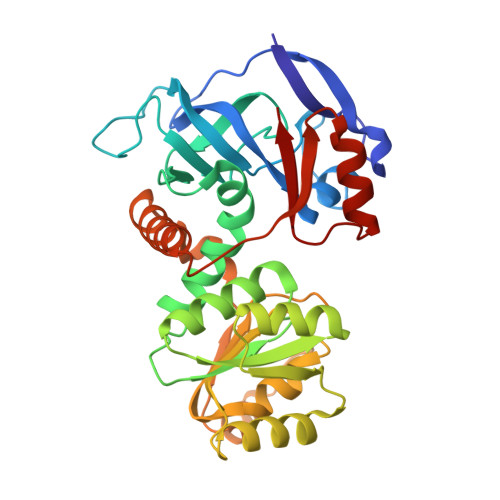
Structural views of quinone oxidoreductase from Mycobacterium tuberculosis reveal large conformational changes induced by the co-factor.
Zheng, Q., Song, Y., Zhang, W., Shaw, N., Zhou, W., Rao, Z.(2015) FEBS J 282: 2697-2707
- PubMed: 25924579
- DOI: https://doi.org/10.1111/febs.13312
- Primary Citation of Related Structures:
4RVS, 4RVU - PubMed Abstract:
Energy generation, synthesis of biomass and detoxification of synthetic compounds are driven by electron transfer in all living organisms. Soluble quinone oxidoreductases (QORs) catalyze transfer of electrons from NADPH to substrates. The open reading frame Rv1454c of Mycobacterium tuberculosis (Mtb) encodes a NADPH-dependent QOR that is known to catalyze one-electron reduction of quinones to produce semiquinones. Here, we report the crystal structures of the apo-enzyme of MtbQOR and its binary complex with NADPH determined at 1.80 and 1.85 Å resolutions, respectively. The enzyme is bi-modular. Domain I binds the substrate, while domain II folds into a typical Rossmann fold for tethering NADPH. Binding of NADPH induces conformational changes. Among the known structures of QORs, MtbQOR exhibits the largest conformational change. Movement of Phe41 to stack against Ala244 results in partial closure of the active site. Comparison of the structure with homologs suggests a conserved topology. However, differences are observed in the region around the site of hydride transfer, highlighting differences in substrate specificities amongst the homologs. Unliganded as well as NADPH-bound MtbQOR crystallized as a dimer. Dimerization is mediated by homotypic intermolecular interactions involving main chain Cα as well as side-chain atoms of residues. The results of analytical ultracentrifugation analysis revealed that MtbQOR exists as a dimer in solution. Enzymatic assays indicate that MtbQOR prefers 9,10-phenanthrenequinone over 1,4-benzoquinone as a substrate. The ability to reduce quinones probably assists Mtb in detoxification of a range of harmful chemicals encountered in the host during invasion.
Organizational Affiliation:
College of Life Sciences, Nankai University, Tianjin, China.