


Crystal Structure of Uncharacterized Membrane Spanning Protein from Vibrio fischeri
Kim, Y., Tesar, C., Bearden, J., Joachimiak, A., Midwest Center for Structural Genomics (MCSG)To be published.
Experimental Data Snapshot
wwPDB Validation 3D Report Full Report

Entity ID: 1 | |||||
|---|---|---|---|---|---|
| Molecule | Chains | Sequence Length | Organism | Details | Image |
| Hypothetical membrane spanning protein | 220 | Aliivibrio fischeri ES114 | Mutation(s): 0 Gene Names: VF_A0681 | 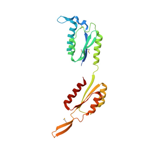 | |
UniProt | |||||
Find proteins for Q5DZP5 (Aliivibrio fischeri (strain ATCC 700601 / ES114)) Explore Q5DZP5 Go to UniProtKB: Q5DZP5 | |||||
Entity Groups | |||||
| Sequence Clusters | 30% Identity50% Identity70% Identity90% Identity95% Identity100% Identity | ||||
| UniProt Group | Q5DZP5 | ||||
Sequence AnnotationsExpand | |||||
| |||||
| Ligands 1 Unique | |||||
|---|---|---|---|---|---|
| ID | Chains | Name / Formula / InChI Key | 2D Diagram | 3D Interactions | |
| MG Query on MG | C [auth A], D [auth B] | MAGNESIUM ION Mg JLVVSXFLKOJNIY-UHFFFAOYSA-N |  | ||
| Modified Residues 1 Unique | |||||
|---|---|---|---|---|---|
| ID | Chains | Type | Formula | 2D Diagram | Parent |
| MSE Query on MSE | A, B | L-PEPTIDE LINKING | C5 H11 N O2 Se |  | MET |
| Length ( Å ) | Angle ( ˚ ) |
|---|---|
| a = 49.734 | α = 90 |
| b = 49.734 | β = 90 |
| c = 343.88 | γ = 120 |
| Software Name | Purpose |
|---|---|
| SBC-Collect | data collection |
| HKL-3000 | data collection |
| HKL-3000 | phasing |
| SHELXS | phasing |
| MLPHARE | phasing |
| DM | model building |
| PHENIX | refinement |
| HKL-3000 | data reduction |
| HKL-3000 | data scaling |
| DM | phasing |
RCSB PDB (citation) is hosted by
RCSB PDB is a member of the

