4RA3

Crystal structure of dimeric S33C beta-2 microglobulin mutant in complex with Thioflavin (ThT) at 2.8 Angstrom resolution


Experimental Data Snapshot

  • Method: X-RAY DIFFRACTION
  • Resolution: 2.80 Å
  • R-Value Free: 0.206 
  • R-Value Work: 0.171 
  • R-Value Observed: 0.173 

wwPDB Validation   3D Report Full Report


Ligand Structure Quality Assessment 


This is version 1.4 of the entry. See complete history


Literature

A covalent homodimer probing early oligomers along amyloid aggregation.

Halabelian, L.Relini, A.Barbiroli, A.Penco, A.Bolognesi, M.Ricagno, S.

(2015) Sci Rep 5: 14651-14651

  • DOI: https://doi.org/10.1038/srep14651
  • Primary Citation of Related Structures:  
    4R9H, 4RA3, 4RAH

  • PubMed Abstract: 

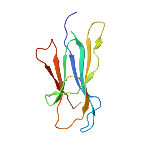
    Early oligomers are crucial in amyloid aggregation; however, due to their transient nature they are among the least structurally characterized species. We focused on the amyloidogenic protein beta2-microglobulin (β2m) whose early oligomers are still a matter of debate. An intermolecular interaction between D strands of facing β2m molecules was repeatedly observed, suggesting that such interface may be relevant for β2m dimerization. In this study, by mutating Ser33 to Cys, and assembling the disulphide-stabilized β2m homodimer (DimC33), such DD strand interface was locked. Although the isolated DimC33 display a stability similar to wt β2m under native conditions, it shows enhanced amyloid aggregation propensity. Three distinct crystal structures of DimC33 suggest that dimerization through the DD interface is instrumental for enhancing DimC33 aggregation propensity. Furthermore, the crystal structure of DimC33 in complex with the amyloid-specific dye Thioflavin-T pinpoints a second interface, which likely participates in the first steps of β2m aggregation. The present data provide new insight into β2m early steps of amyloid aggregation.


  • Organizational Affiliation

    Dipartimento di Bioscienze, Università degli Studi di Milano, Via Celoria 26, 20133 Milan, Italy.


Macromolecules
Find similar proteins by:  (by identity cutoff)  |  3D Structure
Entity ID: 1
MoleculeChains Sequence LengthOrganismDetailsImage
Beta-2-microglobulin
A, B, C, D
100Homo sapiensMutation(s): 1 
Gene Names: B2MCDABP0092HDCMA22PNM_004048
UniProt & NIH Common Fund Data Resources
Find proteins for P61769 (Homo sapiens)
Explore P61769 
Go to UniProtKB:  P61769
PHAROS:  P61769
GTEx:  ENSG00000166710 
Entity Groups  
Sequence Clusters30% Identity50% Identity70% Identity90% Identity95% Identity100% Identity
UniProt GroupP61769
Sequence Annotations
Expand
  • Reference Sequence
Experimental Data & Validation

Experimental Data

  • Method: X-RAY DIFFRACTION
  • Resolution: 2.80 Å
  • R-Value Free: 0.206 
  • R-Value Work: 0.171 
  • R-Value Observed: 0.173 
  • Space Group: P 32 2 1
Unit Cell:
Length ( Å )Angle ( ˚ )
a = 80.038α = 90
b = 80.038β = 90
c = 177.7γ = 120
Software Package:
Software NamePurpose
MxCuBEdata collection
BALBESphasing
REFMACrefinement
XDSdata reduction
SCALAdata scaling

Structure Validation

View Full Validation Report


Ligand Structure Quality Assessment 


Entry History 

Deposition Data

Revision History  (Full details and data files)

  • Version 1.0: 2015-09-09
    Type: Initial release
  • Version 1.1: 2015-09-23
    Changes: Database references
  • Version 1.2: 2015-11-04
    Changes: Database references
  • Version 1.3: 2018-03-07
    Changes: Data collection
  • Version 1.4: 2023-09-20
    Changes: Data collection, Database references, Derived calculations, Refinement description