4R30

Structure of human laforin dual specificity phosphatase domain


Experimental Data Snapshot

  • Method: X-RAY DIFFRACTION
  • Resolution: 2.30 Å
  • R-Value Free: 0.219 
  • R-Value Work: 0.188 
  • R-Value Observed: 0.189 

wwPDB Validation   3D Report Full Report


This is version 1.3 of the entry. See complete history


Literature

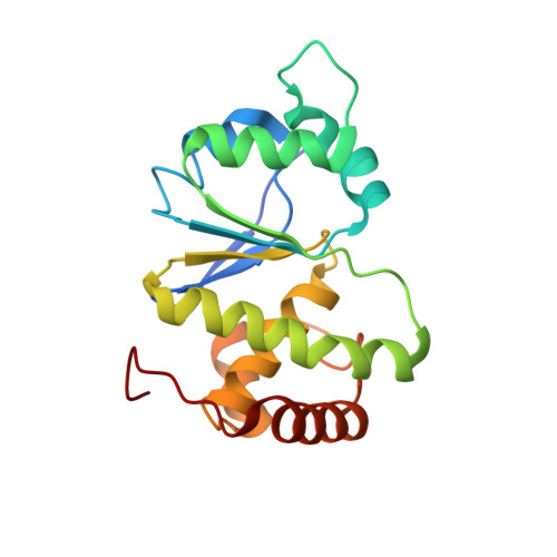
Dimeric quaternary structure of human laforin.

Sankhala, R.S.Koksal, A.C.Ho, L.Nitschke, F.Minassian, B.A.Cingolani, G.

(2015) J Biol Chem 290: 4552-4559

  • DOI: https://doi.org/10.1074/jbc.M114.627406
  • Primary Citation of Related Structures:  
    4R30

  • PubMed Abstract: 

    The phosphatase laforin removes phosphate groups from glycogen during biosynthetic activity. Loss-of-function mutations in the gene encoding laforin is the predominant cause of Lafora disease, a fatal form of progressive myoclonic epilepsy. Here, we used hybrid structural methods to determine the molecular architecture of human laforin. We found that laforin adopts a dimeric quaternary structure, topologically similar to the prototypical dual specificity phosphatase VH1. The interface between the laforin carbohydrate-binding module and the dual specificity phosphatase domain generates an intimate substrate-binding crevice that allows for recognition and dephosphorylation of phosphomonoesters of glucose. We identify novel molecular determinants in the laforin active site that help decipher the mechanism of glucan phosphatase activity.


  • Organizational Affiliation

    From the Department of Biochemistry and Molecular Biology, Thomas Jefferson University, Philadelphia, Pennsylvania 19107.


Macromolecules
Find similar proteins by:  (by identity cutoff)  |  3D Structure
Entity ID: 1
MoleculeChains Sequence LengthOrganismDetailsImage
Laforin
A, B, C, D
184Homo sapiensMutation(s): 1 
Gene Names: EPM2A
EC: 3.1.3 (PDB Primary Data), 3.1.3.16 (PDB Primary Data), 3.1.3.48 (PDB Primary Data)
UniProt & NIH Common Fund Data Resources
Find proteins for O95278 (Homo sapiens)
Explore O95278 
Go to UniProtKB:  O95278
PHAROS:  O95278
GTEx:  ENSG00000112425 
Entity Groups  
Sequence Clusters30% Identity50% Identity70% Identity90% Identity95% Identity100% Identity
UniProt GroupO95278
Sequence Annotations
Expand
  • Reference Sequence
Small Molecules
Ligands 2 Unique
IDChains Name / Formula / InChI Key2D Diagram3D Interactions
SO4
Query on SO4

Download Ideal Coordinates CCD File 
E [auth A],
I [auth B],
M [auth C],
Q [auth D]
SULFATE ION
O4 S
QAOWNCQODCNURD-UHFFFAOYSA-L
BME
Query on BME

Download Ideal Coordinates CCD File 
F [auth A]
G [auth A]
H [auth A]
J [auth B]
K [auth B]
F [auth A],
G [auth A],
H [auth A],
J [auth B],
K [auth B],
L [auth B],
N [auth C],
O [auth C],
P [auth C],
R [auth D],
S [auth D],
T [auth D]
BETA-MERCAPTOETHANOL
C2 H6 O S
DGVVWUTYPXICAM-UHFFFAOYSA-N
Experimental Data & Validation

Experimental Data

  • Method: X-RAY DIFFRACTION
  • Resolution: 2.30 Å
  • R-Value Free: 0.219 
  • R-Value Work: 0.188 
  • R-Value Observed: 0.189 
  • Space Group: I 41
Unit Cell:
Length ( Å )Angle ( ˚ )
a = 123.974α = 90
b = 123.974β = 90
c = 160.771γ = 90
Software Package:
Software NamePurpose
ADSCdata collection
PHASERphasing
PHENIXrefinement
HKL-2000data reduction
HKL-2000data scaling

Structure Validation

View Full Validation Report


Entry History 

Deposition Data

Revision History  (Full details and data files)

  • Version 1.0: 2014-12-31
    Type: Initial release
  • Version 1.1: 2015-01-07
    Changes: Database references
  • Version 1.2: 2015-03-11
    Changes: Database references
  • Version 1.3: 2023-09-20
    Changes: Data collection, Database references, Derived calculations, Refinement description