Structure-based identification of inositol polyphosphate 1-phosphatase from Entamoeba histolytica
Tarique, K.F., Abdul Rehman, S.A., Betzel, C., Gourinath, S.(2014) Acta Crystallogr D Biol Crystallogr 70: 3023-3033
- PubMed: 25372691
- DOI: https://doi.org/10.1107/S1399004714021245
- Primary Citation of Related Structures:
4QXD - PubMed Abstract:
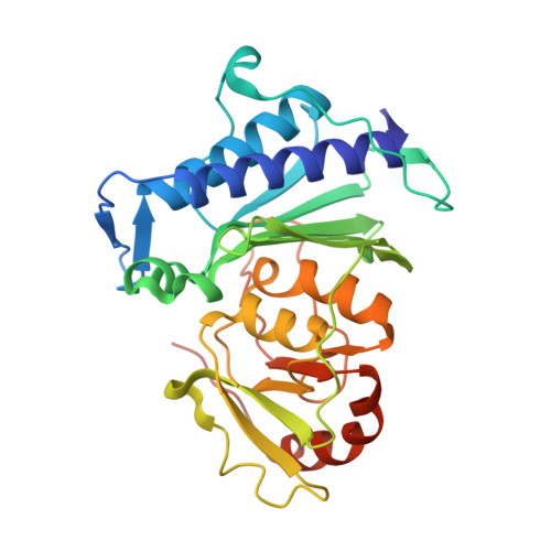
Inositol polyphosphate 1-phosphatase from Entamoeba histolytica (EhIPPase) is an Mg(2+)-dependent and Li(+)-sensitive enzyme that catalyzes the hydrolysis of inositol 1,4-bisphosphate [Ins(1,4)P2] into myo-inositol 1-monophosphate and PO4(3-). In the present work, EhIPPase has been biochemically identified and its crystal structure has been determined in the presence of Mg(2+) and PO4(3-) at 2.5 Å resolution. This enzyme was previously classified as a 3'(2'),5'-bisphosphate nucleotidase in the NCBI, but its biochemical activity and structural analysis suggest that this enzyme behaves more like an inositol polyphosphate 1-phosphatase. The ability of EhIPPase to hydrolyze the smaller Ins(1,4)P2 better than the bulkier 3'-phosphoadenosine 5'-phosphate (PAP) is explained on the basis of the orientations of amino-acid residues in the binding site. This structure is the first of its class to be determined from any protozoan parasite, and is the third to determined among all organisms, following its rat and bovine homologues. The three-dimensional fold of EhIPPase is similar to those of other members of the inositol monophosphatase superfamily, which also includes inositol monophosphatase, 3'(2'),5'-bisphosphate nucleotidase and fructose-1,6-bisphosphate 1-phosphatase. They all share conserved residues essential for metal binding and substrate hydrolysis, with the motif D-Xn-EE-Xn-DP(I/L)DG(S/T)-Xn-WD-Xn-GG. The structure is divided into two domains, namely α+β and α/β, and the substrate and metal ions bind between them. However, the ability of each enzyme class to act specifically on its cognate substrate is governed by the class-specific amino-acid residues at the active site.
Organizational Affiliation:
School of Life Sciences, Jawaharlal Nehru University, New Delhi 110 067, India.