4QOP

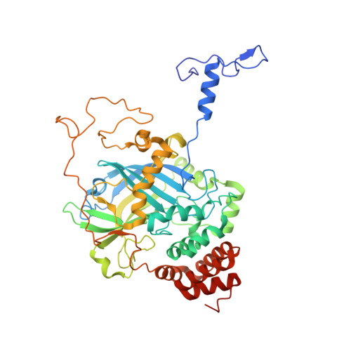
Structure of Bacillus pumilus catalase with hydroquinone bound.


Experimental Data Snapshot

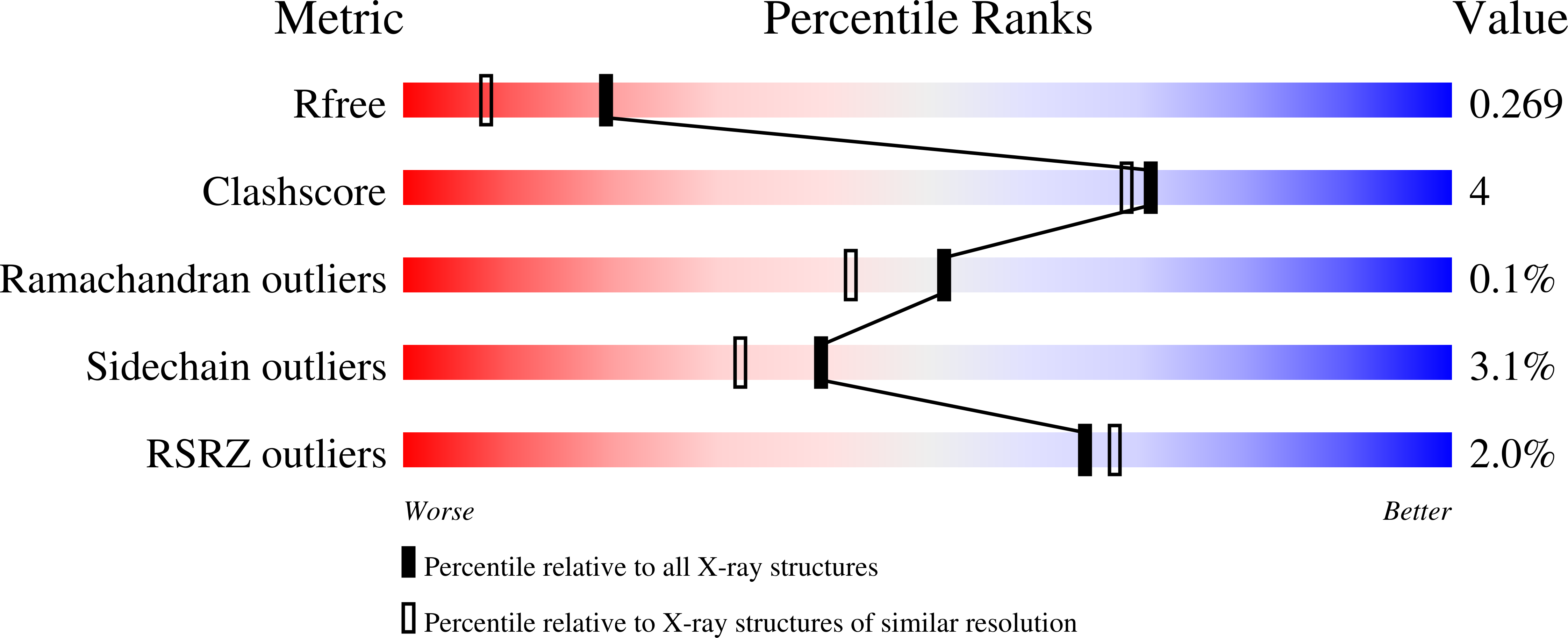
  • Method: X-RAY DIFFRACTION
  • Resolution: 1.90 Å
  • R-Value Free: 0.261 
  • R-Value Work: 0.230 
  • R-Value Observed: 0.231 

wwPDB Validation   3D Report Full Report


Ligand Structure Quality Assessment 


This is version 1.2 of the entry. See complete history


Literature

Unprecedented access of phenolic substrates to the heme active site of a catalase: Substrate binding and peroxidase-like reactivity of Bacillus pumilus catalase monitored by X-ray crystallography and EPR spectroscopy.

Loewen, P.C.Villanueva, J.Switala, J.Donald, L.J.Ivancich, A.

(2015) Proteins 83: 853-866

  • DOI: https://doi.org/10.1002/prot.24777
  • Primary Citation of Related Structures:  
    4QOL, 4QOM, 4QON, 4QOO, 4QOP, 4QOQ, 4QOR

  • PubMed Abstract: 

    Heme-containing catalases and catalase-peroxidases catalyze the dismutation of hydrogen peroxide as their predominant catalytic activity, but in addition, individual enzymes support low levels of peroxidase and oxidase activities, produce superoxide, and activate isoniazid as an antitubercular drug. The recent report of a heme enzyme with catalase, peroxidase and penicillin oxidase activities in Bacillus pumilus and its categorization as an unusual catalase-peroxidase led us to investigate the enzyme for comparison with other catalase-peroxidases, catalases, and peroxidases. Characterization revealed a typical homotetrameric catalase with one pentacoordinated heme b per subunit (Tyr340 being the axial ligand), albeit in two orientations, and a very fast catalatic turnover rate (kcat  = 339,000 s(-1) ). In addition, the enzyme supported a much slower (kcat  = 20 s(-1) ) peroxidatic activity utilizing substrates as diverse as ABTS and polyphenols, but no oxidase activity. Two binding sites, one in the main access channel and the other on the protein surface, accommodating pyrogallol, catechol, resorcinol, guaiacol, hydroquinone, and 2-chlorophenol were identified in crystal structures at 1.65-1.95 Å. A third site, in the heme distal side, accommodating only pyrogallol and catechol, interacting with the heme iron and the catalytic His and Arg residues, was also identified. This site was confirmed in solution by EPR spectroscopy characterization, which also showed that the phenolic oxygen was not directly coordinated to the heme iron (no low-spin conversion of the Fe(III) high-spin EPR signal upon substrate binding). This is the first demonstration of phenolic substrates directly accessing the heme distal side of a catalase.


  • Organizational Affiliation

    Department of Microbiology, University of Manitoba, Winnipeg, Manitoba, R3T 2N2, Canada.


Macromolecules
Find similar proteins by:  (by identity cutoff)  |  3D Structure
Entity ID: 1
MoleculeChains Sequence LengthOrganismDetailsImage
Catalase
A, B, C, D
491Bacillus pumilusMutation(s): 0 
Gene Names: BW16_04845katX2
EC: 1.11.1.6
Entity Groups  
Sequence Clusters30% Identity50% Identity70% Identity90% Identity95% Identity100% Identity
Sequence Annotations
Expand
  • Reference Sequence
Small Molecules
Ligands 4 Unique
IDChains Name / Formula / InChI Key2D Diagram3D Interactions
HEM
Query on HEM

Download Ideal Coordinates CCD File 
E [auth A]
F [auth A]
K [auth B]
L [auth B]
P [auth C]
E [auth A],
F [auth A],
K [auth B],
L [auth B],
P [auth C],
Q [auth C],
U [auth D],
V [auth D]
PROTOPORPHYRIN IX CONTAINING FE
C34 H32 Fe N4 O4
KABFMIBPWCXCRK-RGGAHWMASA-L
HQE
Query on HQE

Download Ideal Coordinates CCD File 
G [auth A],
M [auth B],
R [auth C],
W [auth D]
benzene-1,4-diol
C6 H6 O2
QIGBRXMKCJKVMJ-UHFFFAOYSA-N
CL
Query on CL

Download Ideal Coordinates CCD File 
H [auth A]
I [auth A]
N [auth B]
S [auth C]
T [auth C]
H [auth A],
I [auth A],
N [auth B],
S [auth C],
T [auth C],
X [auth D]
CHLORIDE ION
Cl
VEXZGXHMUGYJMC-UHFFFAOYSA-M
NA
Query on NA

Download Ideal Coordinates CCD File 
J [auth A],
O [auth B]
SODIUM ION
Na
FKNQFGJONOIPTF-UHFFFAOYSA-N
Experimental Data & Validation

Experimental Data

  • Method: X-RAY DIFFRACTION
  • Resolution: 1.90 Å
  • R-Value Free: 0.261 
  • R-Value Work: 0.230 
  • R-Value Observed: 0.231 
  • Space Group: P 1 21 1
Unit Cell:
Length ( Å )Angle ( ˚ )
a = 92.279α = 90
b = 109.455β = 92.05
c = 104.199γ = 90
Software Package:
Software NamePurpose
SCALAdata scaling
REFMACrefinement
PDB_EXTRACTdata extraction
XDSdata scaling
XDSdata reduction
MOLREPphasing

Structure Validation

View Full Validation Report


Ligand Structure Quality Assessment 


Entry History 

Deposition Data

  • Released Date: 2015-02-25 
  • Deposition Author(s): Loewen, P.C.

Revision History  (Full details and data files)

  • Version 1.0: 2015-02-25
    Type: Initial release
  • Version 1.1: 2015-04-29
    Changes: Database references
  • Version 1.2: 2024-02-28
    Changes: Data collection, Database references, Derived calculations, Refinement description