4QHO

Crystal structure of the human fat mass and obesity associated protein (FTO) in complex with CCO10


Experimental Data Snapshot

  • Method: X-RAY DIFFRACTION
  • Resolution: 2.37 Å
  • R-Value Free: 0.208 
  • R-Value Work: 0.177 

wwPDB Validation   3D Report Full Report


Ligand Structure Quality Assessment 


This is version 1.3 of the entry. See complete history


Literature

Structure-Based Design of Selective Fat Mass and Obesity Associated Protein (FTO) Inhibitors.

Shishodia, S.Demetriades, M.Zhang, D.Tam, N.Y.Maheswaran, P.Clunie-O'Connor, C.Tumber, A.Leung, I.K.H.Ng, Y.M.Leissing, T.M.El-Sagheer, A.H.Salah, E.Brown, T.Aik, W.S.McDonough, M.A.Schofield, C.J.

(2021) J Med Chem 64: 16609-16625

  • DOI: https://doi.org/10.1021/acs.jmedchem.1c01204
  • Primary Citation of Related Structures:  
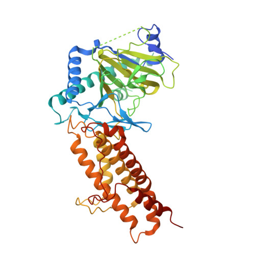
    4QHO, 7E8Z, 7NRO

  • PubMed Abstract: 

    FTO catalyzes the Fe(II) and 2-oxoglutarate (2OG)-dependent modification of nucleic acids, including the demethylation of N 6 -methyladenosine (m 6 A) in mRNA. FTO is a proposed target for anti-cancer therapy. Using information from crystal structures of FTO in complex with 2OG and substrate mimics, we designed and synthesized two series of FTO inhibitors, which were characterized by turnover and binding assays, and by X-ray crystallography with FTO and the related bacterial enzyme AlkB. A potent inhibitor employing binding interactions spanning the FTO 2OG and substrate binding sites was identified. Selectivity over other clinically targeted 2OG oxygenases was demonstrated, including with respect to the hypoxia-inducible factor prolyl and asparaginyl hydroxylases (PHD2 and FIH) and selected JmjC histone demethylases (KDMs). The results illustrate how structure-based design can enable the identification of potent and selective 2OG oxygenase inhibitors and will be useful for the development of FTO inhibitors for use in vivo .


  • Organizational Affiliation

    The Chemistry Research Laboratory, Department of Chemistry and the Ineos Oxford Institute for Antimicrobial Research, University of Oxford, 12 Mansfield Road, Oxford OX1 3TA, U.K.


Macromolecules
Find similar proteins by:  (by identity cutoff)  |  3D Structure
Entity ID: 1
MoleculeChains Sequence LengthOrganismDetailsImage
Alpha-ketoglutarate-dependent dioxygenase FTO495Homo sapiensMutation(s): 0 
Gene Names: FTOKIAA1752
EC: 1.14.11
UniProt & NIH Common Fund Data Resources
Find proteins for Q9C0B1 (Homo sapiens)
Explore Q9C0B1 
Go to UniProtKB:  Q9C0B1
PHAROS:  Q9C0B1
GTEx:  ENSG00000140718 
Entity Groups  
Sequence Clusters30% Identity50% Identity70% Identity90% Identity95% Identity100% Identity
UniProt GroupQ9C0B1
Sequence Annotations
Expand
  • Reference Sequence
Small Molecules
Ligands 2 Unique
IDChains Name / Formula / InChI Key2D Diagram3D Interactions
NKG
Query on NKG

Download Ideal Coordinates CCD File 
C [auth A]N-{[3-hydroxy-6-(naphthalen-1-yl)pyridin-2-yl]carbonyl}glycine
C18 H14 N2 O4
CQPTZSBTFPQERZ-UHFFFAOYSA-N
ZN
Query on ZN

Download Ideal Coordinates CCD File 
B [auth A]ZINC ION
Zn
PTFCDOFLOPIGGS-UHFFFAOYSA-N
Experimental Data & Validation

Experimental Data

  • Method: X-RAY DIFFRACTION
  • Resolution: 2.37 Å
  • R-Value Free: 0.208 
  • R-Value Work: 0.177 
  • Space Group: H 3
Unit Cell:
Length ( Å )Angle ( ˚ )
a = 141.384α = 90
b = 141.384β = 90
c = 83.954γ = 120
Software Package:
Software NamePurpose
DENZOdata reduction
SCALEPACKdata scaling
PHENIXrefinement
PDB_EXTRACTdata extraction
GDAdata collection
HKL-3000data reduction
HKL-3000data scaling
PHASERphasing

Structure Validation

View Full Validation Report


Ligand Structure Quality Assessment 


Entry History 

Deposition Data

Revision History  (Full details and data files)

  • Version 1.0: 2015-06-03
    Type: Initial release
  • Version 1.1: 2021-10-13
    Changes: Database references, Derived calculations, Structure summary
  • Version 1.2: 2022-02-02
    Changes: Database references
  • Version 1.3: 2023-11-08
    Changes: Data collection, Refinement description