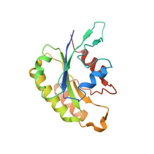
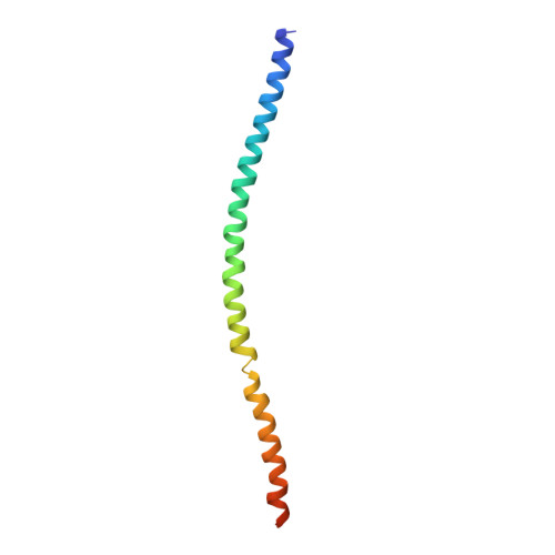
4Q9U

Crystal structure of the Rab5, Rabex-5delta and Rabaptin-5C21 complex


Experimental Data Snapshot

  • Method: X-RAY DIFFRACTION
  • Resolution: 4.62 Å
  • R-Value Free: 0.343 
  • R-Value Work: 0.251 
  • R-Value Observed: 0.256 

wwPDB Validation   3D Report Full Report


This is version 1.3 of the entry. See complete history


Literature

Molecular mechanism for Rabex-5 GEF activation by Rabaptin-5

Zhang, Z.Zhang, T.Wang, S.Gong, Z.Tang, C.Chen, J.Ding, J.

(2014) Elife 3: e02687-e02687

  • DOI: https://doi.org/10.7554/eLife.02687
  • Primary Citation of Related Structures:  
    4N3X, 4N3Y, 4N3Z, 4Q9U

  • PubMed Abstract: 

    Rabex-5 and Rabaptin-5 function together to activate Rab5 and further promote early endosomal fusion in endocytosis. The Rabex-5 GEF activity is autoinhibited by the Rabex-5 CC domain (Rabex-5CC) and activated by the Rabaptin-5 C2-1 domain (Rabaptin-5C21) with yet unknown mechanism. We report here the crystal structures of Rabex-5 in complex with the dimeric Rabaptin-5C21 (Rabaptin-5C212) and in complex with Rabaptin-5C212 and Rab5, along with biophysical and biochemical analyses. We show that Rabex-5CC assumes an amphipathic α-helix which binds weakly to the substrate-binding site of the GEF domain, leading to weak autoinhibition of the GEF activity. Binding of Rabaptin-5C21 to Rabex-5 displaces Rabex-5CC to yield a largely exposed substrate-binding site, leading to release of the GEF activity. In the ternary complex the substrate-binding site of Rabex-5 is completely exposed to bind and activate Rab5. Our results reveal the molecular mechanism for the regulation of the Rabex-5 GEF activity.


  • Organizational Affiliation

    State Key Laboratory of Molecular Biology, Institute of Biochemistry and Cell Biology, Shanghai Institutes for Biological Sciences, Chinese Academy of Sciences, Shanghai, China.


Macromolecules
Find similar proteins by:  (by identity cutoff)  |  3D Structure
Entity ID: 1
MoleculeChains Sequence LengthOrganismDetailsImage
Rab5 GDP/GTP exchange factor
A, E
317Homo sapiensMutation(s): 0 
Gene Names: RABGEF1RABEX5
UniProt & NIH Common Fund Data Resources
Find proteins for Q9UJ41 (Homo sapiens)
Explore Q9UJ41 
Go to UniProtKB:  Q9UJ41
GTEx:  ENSG00000154710 
Entity Groups  
Sequence Clusters30% Identity50% Identity70% Identity90% Identity95% Identity100% Identity
UniProt GroupQ9UJ41
Sequence Annotations
Expand
  • Reference Sequence
Find similar proteins by:  (by identity cutoff)  |  3D Structure
Entity ID: 2
MoleculeChains Sequence LengthOrganismDetailsImage
Ras-related protein Rab-5A
B, F
171Homo sapiensMutation(s): 0 
Gene Names: RAB5ARAB5
UniProt & NIH Common Fund Data Resources
Find proteins for P20339 (Homo sapiens)
Explore P20339 
Go to UniProtKB:  P20339
PHAROS:  P20339
GTEx:  ENSG00000144566 
Entity Groups  
Sequence Clusters30% Identity50% Identity70% Identity90% Identity95% Identity100% Identity
UniProt GroupP20339
Sequence Annotations
Expand
  • Reference Sequence
Find similar proteins by:  (by identity cutoff)  |  3D Structure
Entity ID: 3
MoleculeChains Sequence LengthOrganismDetailsImage
Rab GTPase-binding effector protein 1
C, D, G, H
92Homo sapiensMutation(s): 0 
Gene Names: RABEP1RAB5EPRABPT5RABPT5A
UniProt & NIH Common Fund Data Resources
Find proteins for Q15276 (Homo sapiens)
Explore Q15276 
Go to UniProtKB:  Q15276
PHAROS:  Q15276
GTEx:  ENSG00000029725 
Entity Groups  
Sequence Clusters30% Identity50% Identity70% Identity90% Identity95% Identity100% Identity
UniProt GroupQ15276
Sequence Annotations
Expand
  • Reference Sequence
Experimental Data & Validation

Experimental Data

  • Method: X-RAY DIFFRACTION
  • Resolution: 4.62 Å
  • R-Value Free: 0.343 
  • R-Value Work: 0.251 
  • R-Value Observed: 0.256 
  • Space Group: P 41 21 2
Unit Cell:
Length ( Å )Angle ( ˚ )
a = 174.821α = 90
b = 174.821β = 90
c = 148.995γ = 90
Software Package:
Software NamePurpose
DENZOdata reduction
SCALEPACKdata scaling
PHASERphasing
PHENIXrefinement
PDB_EXTRACTdata extraction
HKL-2000data collection

Structure Validation

View Full Validation Report


Entry History 

Deposition Data

Revision History  (Full details and data files)

  • Version 1.0: 2014-07-23
    Type: Initial release
  • Version 1.1: 2015-12-09
    Changes: Database references
  • Version 1.2: 2017-11-22
    Changes: Refinement description
  • Version 1.3: 2023-11-08
    Changes: Data collection, Database references, Refinement description