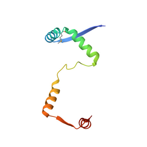
4Q2U

Crystal structure of the E. coli DinJ-YafQ toxin-antitoxin complex


Experimental Data Snapshot

  • Method: X-RAY DIFFRACTION
  • Resolution: 1.80 Å
  • R-Value Free: 0.215 
  • R-Value Work: 0.178 
  • R-Value Observed: 0.178 

wwPDB Validation   3D Report Full Report


This is version 1.2 of the entry. See complete history


Literature

Mechanisms of Toxin Inhibition and Transcriptional Repression by Escherichia coli DinJ-YafQ.

Ruangprasert, A.Maehigashi, T.Miles, S.J.Giridharan, N.Liu, J.X.Dunham, C.M.

(2014) J Biol Chem 289: 20559-20569

  • DOI: https://doi.org/10.1074/jbc.M114.573006
  • Primary Citation of Related Structures:  
    4Q2U

  • PubMed Abstract: 

    Bacteria encounter environmental stresses that regulate a gene expression program required for adaptation and survival. Here, we report the 1.8-Å crystal structure of the Escherichia coli toxin-antitoxin complex YafQ-(DinJ)2-YafQ, a key component of the stress response. The antitoxin DinJ dimer adopts a ribbon-helix-helix motif required for transcriptional autorepression, and toxin YafQ contains a microbial RNase fold whose proposed active site is concealed by DinJ binding. Contrary to previous reports, our studies indicate that equivalent levels of transcriptional repression occur by direct interaction of either YafQ-(DinJ)2-YafQ or a DinJ dimer at a single inverted repeat of its recognition sequence that overlaps with the -10 promoter region. Surprisingly, multiple YafQ-(DinJ)2-YafQ complexes binding to the operator region do not appear to amplify the extent of repression. Our results suggest an alternative model for transcriptional autorepression that may be novel to DinJ-YafQ.


  • Organizational Affiliation

    From the Department of Biochemistry, Emory University School of Medicine, Atlanta, Georgia 30322.


Macromolecules
Find similar proteins by:  (by identity cutoff)  |  3D Structure
Entity ID: 1
MoleculeChains Sequence LengthOrganismDetailsImage
Antitoxin DinJ
A, C, E, G, I
A, C, E, G, I, K, M, O
86Escherichia coli K-12Mutation(s): 0 
Gene Names: dinJb0226JW0216
UniProt
Find proteins for Q47150 (Escherichia coli (strain K12))
Explore Q47150 
Go to UniProtKB:  Q47150
Entity Groups  
Sequence Clusters30% Identity50% Identity70% Identity90% Identity95% Identity100% Identity
UniProt GroupQ47150
Sequence Annotations
Expand
  • Reference Sequence
Find similar proteins by:  (by identity cutoff)  |  3D Structure
Entity ID: 2
MoleculeChains Sequence LengthOrganismDetailsImage
mRNA interferase YafQ
B, D, F, H, J
B, D, F, H, J, L, N, P
102Escherichia coli K-12Mutation(s): 0 
Gene Names: yafQb0225JW0215
EC: 3.1
UniProt
Find proteins for Q47149 (Escherichia coli (strain K12))
Explore Q47149 
Go to UniProtKB:  Q47149
Entity Groups  
Sequence Clusters30% Identity50% Identity70% Identity90% Identity95% Identity100% Identity
UniProt GroupQ47149
Sequence Annotations
Expand
  • Reference Sequence
Small Molecules
Ligands 1 Unique
IDChains Name / Formula / InChI Key2D Diagram3D Interactions
SO4
Query on SO4

Download Ideal Coordinates CCD File 
AA [auth C]
AB [auth H]
BA [auth C]
BB [auth I]
CA [auth C]
AA [auth C],
AB [auth H],
BA [auth C],
BB [auth I],
CA [auth C],
CB [auth J],
DA [auth C],
DB [auth J],
EA [auth D],
EB [auth K],
FA [auth D],
FB [auth L],
GA [auth D],
GB [auth L],
HA [auth D],
HB [auth M],
IA [auth D],
IB [auth N],
JA [auth E],
JB [auth N],
KA [auth E],
KB [auth O],
LA [auth E],
LB [auth P],
MA [auth F],
MB [auth P],
NA [auth F],
OA [auth F],
PA [auth F],
Q [auth A],
QA [auth F],
R [auth A],
RA [auth G],
S [auth A],
SA [auth G],
T [auth A],
TA [auth G],
U [auth B],
UA [auth G],
V [auth B],
VA [auth H],
W [auth B],
WA [auth H],
X [auth B],
XA [auth H],
Y [auth B],
YA [auth H],
Z [auth B],
ZA [auth H]
SULFATE ION
O4 S
QAOWNCQODCNURD-UHFFFAOYSA-L
Modified Residues  1 Unique
IDChains TypeFormula2D DiagramParent
MSE
Query on MSE
A, C, E, G, I
A, C, E, G, I, K, M, O
L-PEPTIDE LINKINGC5 H11 N O2 SeMET
Experimental Data & Validation

Experimental Data

  • Method: X-RAY DIFFRACTION
  • Resolution: 1.80 Å
  • R-Value Free: 0.215 
  • R-Value Work: 0.178 
  • R-Value Observed: 0.178 
  • Space Group: C 1 2 1
Unit Cell:
Length ( Å )Angle ( ˚ )
a = 176.54α = 90
b = 120.92β = 130.77
c = 120.83γ = 90
Software Package:
Software NamePurpose
SHELXDphasing
PHENIXmodel building
PHENIXrefinement
HKL-2000data reduction
HKL-2000data scaling
PHENIXphasing

Structure Validation

View Full Validation Report


Entry History 

Deposition Data

Revision History  (Full details and data files)

  • Version 1.0: 2014-06-11
    Type: Initial release
  • Version 1.1: 2014-07-02
    Changes: Database references
  • Version 1.2: 2014-08-06
    Changes: Database references