4PE5

Crystal Structure of GluN1a/GluN2B NMDA Receptor Ion Channel

  • Classification: TRANSPORT PROTEIN
  • Organism(s): Rattus norvegicus
  • Expression System: Spodoptera frugiperda
  • Mutation(s): Yes 
  • Membrane Protein: Yes  OPMPDBTMMemProtMD

  • Deposited: 2014-04-22 Released: 2014-06-04 
  • Deposition Author(s): Karakas, E., Furukawa, H.
  • Funding Organization(s): National Institutes of Health/National Institute of General Medical Sciences (NIH/NIGMS), National Institutes of Health/National Institute of Mental Health (NIH/NIMH), Robertson Research Fund, Mirus Research Award

Experimental Data Snapshot

  • Method: X-RAY DIFFRACTION
  • Resolution: 3.96 Å
  • R-Value Free: 0.295 
  • R-Value Work: 0.256 
  • R-Value Observed: 0.258 

wwPDB Validation   3D Report Full Report


This is version 3.1 of the entry. See complete history


Literature

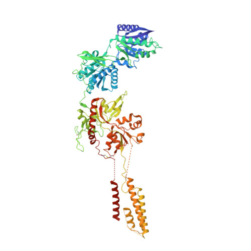
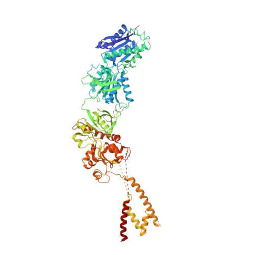
Crystal structure of a heterotetrameric NMDA receptor ion channel.

Karakas, E.Furukawa, H.

(2014) Science 344: 992-997

  • DOI: https://doi.org/10.1126/science.1251915
  • Primary Citation of Related Structures:  
    4PE5

  • PubMed Abstract: 

    N-Methyl-D-aspartate (NMDA) receptors belong to the family of ionotropic glutamate receptors, which mediate most excitatory synaptic transmission in mammalian brains. Calcium permeation triggered by activation of NMDA receptors is the pivotal event for initiation of neuronal plasticity. Here, we show the crystal structure of the intact heterotetrameric GluN1-GluN2B NMDA receptor ion channel at 4 angstroms. The NMDA receptors are arranged as a dimer of GluN1-GluN2B heterodimers with the twofold symmetry axis running through the entire molecule composed of an amino terminal domain (ATD), a ligand-binding domain (LBD), and a transmembrane domain (TMD). The ATD and LBD are much more highly packed in the NMDA receptors than non-NMDA receptors, which may explain why ATD regulates ion channel activity in NMDA receptors but not in non-NMDA receptors.


  • Organizational Affiliation

    Cold Spring Harbor Laboratory, W. M. Keck Structural Biology Laboratory, One Bungtown Road, Cold Spring Harbor, NY 11724, USA.


Macromolecules
Find similar proteins by:  (by identity cutoff)  |  3D Structure
Entity ID: 1
MoleculeChains Sequence LengthOrganismDetailsImage
Glutamate receptor ionotropic, NMDA 1
A, C
825Rattus norvegicusMutation(s): 15 
Gene Names: Grin1Nmdar1
Membrane Entity: Yes 
UniProt
Find proteins for P35439 (Rattus norvegicus)
Explore P35439 
Go to UniProtKB:  P35439
Entity Groups  
Sequence Clusters30% Identity50% Identity70% Identity90% Identity95% Identity100% Identity
UniProt GroupP35439
Sequence Annotations
Expand
  • Reference Sequence
Find similar proteins by:  (by identity cutoff)  |  3D Structure
Entity ID: 2
MoleculeChains Sequence LengthOrganismDetailsImage
Glutamate receptor ionotropic, NMDA 2B
B, D
820Rattus norvegicusMutation(s): 7 
Gene Names: Grin2b
Membrane Entity: Yes 
UniProt
Find proteins for Q00960 (Rattus norvegicus)
Explore Q00960 
Go to UniProtKB:  Q00960
Entity Groups  
Sequence Clusters30% Identity50% Identity70% Identity90% Identity95% Identity100% Identity
UniProt GroupQ00960
Sequence Annotations
Expand
  • Reference Sequence
Oligosaccharides

Help

Entity ID: 3
MoleculeChains Length2D Diagram Glycosylation3D Interactions
alpha-D-mannopyranose-(1-6)-beta-D-mannopyranose-(1-4)-2-acetamido-2-deoxy-beta-D-glucopyranose-(1-4)-2-acetamido-2-deoxy-beta-D-glucopyranose
E, I, J
4N-Glycosylation
Glycosylation Resources
GlyTouCan:  G22573RC
GlyCosmos:  G22573RC
GlyGen:  G22573RC
Entity ID: 4
MoleculeChains Length2D Diagram Glycosylation3D Interactions
alpha-D-mannopyranose-(1-3)-alpha-D-mannopyranose-(1-6)-[alpha-D-mannopyranose-(1-3)]beta-D-mannopyranose-(1-4)-2-acetamido-2-deoxy-beta-D-glucopyranose-(1-4)-2-acetamido-2-deoxy-beta-D-glucopyranose
F
6N-Glycosylation
Glycosylation Resources
GlyTouCan:  G09724ZC
GlyCosmos:  G09724ZC
GlyGen:  G09724ZC
Entity ID: 5
MoleculeChains Length2D Diagram Glycosylation3D Interactions
alpha-D-mannopyranose-(1-6)-alpha-D-mannopyranose-(1-6)-alpha-D-mannopyranose-(1-6)-beta-D-mannopyranose-(1-4)-2-acetamido-2-deoxy-beta-D-glucopyranose-(1-4)-2-acetamido-2-deoxy-beta-D-glucopyranose
G
6N-Glycosylation
Glycosylation Resources
GlyTouCan:  G76865YT
GlyCosmos:  G76865YT
GlyGen:  G76865YT
Entity ID: 6
MoleculeChains Length2D Diagram Glycosylation3D Interactions
2-acetamido-2-deoxy-beta-D-glucopyranose-(1-4)-2-acetamido-2-deoxy-beta-D-glucopyranose
H
2N-Glycosylation
Glycosylation Resources
GlyTouCan:  G42666HT
GlyCosmos:  G42666HT
GlyGen:  G42666HT
Small Molecules
Ligands 5 Unique
IDChains Name / Formula / InChI Key2D Diagram3D Interactions
QEL
Query on QEL

Download Ideal Coordinates CCD File 
MB [auth B],
YC [auth C]
4-[(1R,2S)-2-(4-benzylpiperidin-1-yl)-1-hydroxypropyl]phenol
C21 H27 N O2
UYNVMODNBIQBMV-KKSFZXQISA-N
NAG
Query on NAG

Download Ideal Coordinates CCD File 
AD [auth C]
BD [auth D]
CD [auth D]
KB [auth B]
LB [auth B]
AD [auth C],
BD [auth D],
CD [auth D],
KB [auth B],
LB [auth B],
WA [auth A],
XA [auth A],
YA [auth A]
2-acetamido-2-deoxy-beta-D-glucopyranose
C8 H15 N O6
OVRNDRQMDRJTHS-FMDGEEDCSA-N
W
Query on W

Download Ideal Coordinates CCD File 
AA [auth A]
AB [auth B]
AC [auth C]
BA [auth A]
BB [auth B]
AA [auth A],
AB [auth B],
AC [auth C],
BA [auth A],
BB [auth B],
BC [auth C],
CA [auth A],
CB [auth B],
CC [auth C],
DA [auth A],
DB [auth B],
DC [auth C],
EA [auth A],
EB [auth B],
EC [auth C],
FA [auth A],
FB [auth B],
FC [auth C],
GA [auth A],
GB [auth B],
GC [auth C],
HA [auth A],
HB [auth B],
HC [auth C],
IA [auth A],
IB [auth B],
IC [auth C],
JA [auth A],
JB [auth B],
JC [auth C],
K [auth A],
KA [auth A],
KC [auth C],
L [auth A],
LA [auth A],
LC [auth C],
M [auth A],
MA [auth A],
MC [auth C],
N [auth A],
NA [auth A],
NC [auth C],
O [auth A],
OA [auth A],
OB [auth C],
OC [auth C],
P [auth A],
PA [auth A],
PB [auth C],
PC [auth C],
Q [auth A],
QA [auth A],
QB [auth C],
QC [auth C],
R [auth A],
RA [auth A],
RB [auth C],
RC [auth C],
S [auth A],
SA [auth A],
SB [auth C],
SC [auth C],
T [auth A],
TA [auth A],
TB [auth C],
TC [auth C],
U [auth A],
UA [auth A],
UB [auth C],
UC [auth C],
V [auth A],
VB [auth C],
VC [auth C],
W [auth A],
WB [auth C],
WC [auth C],
X [auth A],
XB [auth C],
XC [auth C],
Y [auth A],
YB [auth C],
Z [auth A],
ZA [auth B],
ZB [auth C]
TUNGSTEN ION
W
FZFRVZDLZISPFJ-UHFFFAOYSA-N
GLU
Query on GLU

Download Ideal Coordinates CCD File 
DD [auth D],
NB [auth B]
GLUTAMIC ACID
C5 H9 N O4
WHUUTDBJXJRKMK-VKHMYHEASA-N
GLY
Query on GLY

Download Ideal Coordinates CCD File 
VA [auth A],
ZC [auth C]
GLYCINE
C2 H5 N O2
DHMQDGOQFOQNFH-UHFFFAOYSA-N
Binding Affinity Annotations 
IDSourceBinding Affinity
QEL BindingDB:  4PE5 Ki: min: 10, max: 29 (nM) from 6 assay(s)
Kd: min: 7.6, max: 94 (nM) from 2 assay(s)
IC50: min: 20, max: 1.00e+5 (nM) from 13 assay(s)
Experimental Data & Validation

Experimental Data

  • Method: X-RAY DIFFRACTION
  • Resolution: 3.96 Å
  • R-Value Free: 0.295 
  • R-Value Work: 0.256 
  • R-Value Observed: 0.258 
  • Space Group: P 1 21 1
Unit Cell:
Length ( Å )Angle ( ˚ )
a = 116.825α = 90
b = 163.186β = 93.81
c = 163.137γ = 90
Software Package:
Software NamePurpose
PHENIXrefinement
HKL-2000data reduction
HKL-2000data scaling
PDB_EXTRACTdata extraction

Structure Validation

View Full Validation Report


Entry History & Funding Information

Deposition Data


Funding OrganizationLocationGrant Number
National Institutes of Health/National Institute of General Medical Sciences (NIH/NIGMS)United StatesGM105730
National Institutes of Health/National Institute of Mental Health (NIH/NIMH)United StatesMH085926
Robertson Research FundUnited States--
Mirus Research AwardUnited States--

Revision History  (Full details and data files)

  • Version 1.0: 2014-06-04
    Type: Initial release
  • Version 1.1: 2014-06-11
    Changes: Database references
  • Version 1.2: 2014-07-30
    Changes: Derived calculations
  • Version 2.0: 2017-09-06
    Changes: Atomic model, Author supporting evidence, Database references, Derived calculations, Other, Source and taxonomy, Structure summary
  • Version 2.1: 2019-11-27
    Changes: Author supporting evidence
  • Version 3.0: 2020-07-29
    Type: Remediation
    Reason: Carbohydrate remediation
    Changes: Advisory, Atomic model, Data collection, Derived calculations, Refinement description, Structure summary
  • Version 3.1: 2023-09-27
    Changes: Data collection, Database references, Refinement description, Structure summary