Structures of darunavir-resistant HIV-1 protease mutant reveal atypical binding of darunavir to wide open flaps.
Zhang, Y., Chang, Y.C., Louis, J.M., Wang, Y.F., Harrison, R.W., Weber, I.T.(2014) ACS Chem Biol 9: 1351-1358
- PubMed: 24738918
- DOI: https://doi.org/10.1021/cb4008875
- Primary Citation of Related Structures:
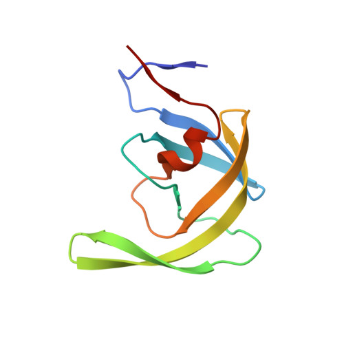
4NPT, 4NPU - PubMed Abstract:
The molecular basis for high resistance to clinical inhibitors of HIV-1 protease (PR) was examined for the variant designated PRP51 that was selected for resistance to darunavir (DRV). High resolution crystal structures of PRP51 with the active site D25N mutation revealed a ligand-free form and an inhibitor-bound form showing a unique binding site and orientation for DRV. This inactivating mutation is known to increase the dimer dissociation constant and decrease DRV affinity of PR. The PRP51-D25N dimers were in the open conformation with widely separated flaps, as reported for other highly resistant variants. PRP51-D25N dimer bound two DRV molecules and showed larger separation of 8.7 Å between the closest atoms of the two flaps compared with 4.4 Å for the ligand-free structure of this mutant. The ligand-free structure, however, lacked van der Waals contacts between Ile50 and Pro81' from the other subunit in the dimer, unlike the majority of PR structures. DRV is bound inside the active site cavity; however, the inhibitor is oriented almost perpendicular to its typical position and exhibits only 2 direct hydrogen bond and two water-mediated interactions with atoms of PRP51-D25N compared with 11 hydrogen bond interactions seen for DRV bound in the typical position in wild-type enzyme. The atypical location of DRV may provide opportunities for design of novel inhibitors targeting the open conformation of PR drug-resistant mutants.
Organizational Affiliation:
Department of Chemistry, ‡Department of Biology, and §Department of Computer Science, Georgia State University , Atlanta, Georgia 30303, United States.