4MV2

Crystal structure of plu4264 protein from Photorhabdus luminescens


Experimental Data Snapshot

  • Method: X-RAY DIFFRACTION
  • Resolution: 1.35 Å
  • R-Value Free: 0.159 
  • R-Value Work: 0.136 
  • R-Value Observed: 0.136 

wwPDB Validation   3D Report Full Report


This is version 1.3 of the entry. See complete history


Literature

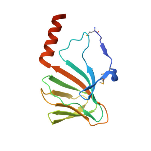
Structure of a cupin protein Plu4264 from Photorhabdus luminescens subsp. laumondii TTO1 at 1.35 angstrom resolution.

Weerth, R.S.Michalska, K.Bingman, C.A.Yennamalli, R.M.Li, H.Jedrzejczak, R.Wang, F.Babnigg, G.Joachimiak, A.Thomas, M.G.Phillips, G.N.

(2015) Proteins 83: 383-388

  • DOI: https://doi.org/10.1002/prot.24705
  • Primary Citation of Related Structures:  
    4MV2, 4Q29

  • PubMed Abstract: 

    Proteins belonging to the cupin superfamily have a wide range of catalytic and noncatalytic functions. Cupin proteins commonly have the capacity to bind a metal ion with the metal frequently determining the function of the protein. We have been investigating the function of homologous cupin proteins that are conserved in more than 40 species of bacteria. To gain insights into the potential function of these proteins we have solved the structure of Plu4264 from Photorhabdus luminescens TTO1 at a resolution of 1.35 Å and identified manganese as the likely natural metal ligand of the protein.


  • Organizational Affiliation

    Department of Bacteriology, University of Wisconsin-Madison, Madison, Wisconsin.


Macromolecules
Find similar proteins by:  (by identity cutoff)  |  3D Structure
Entity ID: 1
MoleculeChains Sequence LengthOrganismDetailsImage
plu4264
A, B
128Photorhabdus laumondii subsp. laumondii TTO1Mutation(s): 0 
Gene Names: plu4264
UniProt
Find proteins for Q7MZL9 (Photorhabdus laumondii subsp. laumondii (strain DSM 15139 / CIP 105565 / TT01))
Explore Q7MZL9 
Go to UniProtKB:  Q7MZL9
Entity Groups  
Sequence Clusters30% Identity50% Identity70% Identity90% Identity95% Identity100% Identity
UniProt GroupQ7MZL9
Sequence Annotations
Expand
  • Reference Sequence
Small Molecules
Modified Residues  1 Unique
IDChains TypeFormula2D DiagramParent
MSE
Query on MSE
A, B
L-PEPTIDE LINKINGC5 H11 N O2 SeMET
Experimental Data & Validation

Experimental Data

  • Method: X-RAY DIFFRACTION
  • Resolution: 1.35 Å
  • R-Value Free: 0.159 
  • R-Value Work: 0.136 
  • R-Value Observed: 0.136 
  • Space Group: C 2 2 21
Unit Cell:
Length ( Å )Angle ( ˚ )
a = 55.738α = 90
b = 147.687β = 90
c = 83.843γ = 90
Software Package:
Software NamePurpose
SBC-Collectdata collection
SHELXmodel building
MLPHAREphasing
DMmodel building
ARP/wARPmodel building
Cootmodel building
PHENIXrefinement
HKL-3000data reduction
HKL-3000data scaling
SHELXphasing
DMphasing

Structure Validation

View Full Validation Report


Entry History 

Revision History  (Full details and data files)

  • Version 1.0: 2013-10-02
    Type: Initial release
  • Version 1.1: 2013-10-09
    Changes: Source and taxonomy
  • Version 1.2: 2014-11-12
    Changes: Database references
  • Version 1.3: 2015-02-04
    Changes: Database references