Enzymatic Basis for N-Glycan Sialylation: STRUCTURE OF RAT alpha 2,6-SIALYLTRANSFERASE (ST6GAL1) REVEALS CONSERVED AND UNIQUE FEATURES FOR GLYCAN SIALYLATION.
Meng, L., Forouhar, F., Thieker, D., Gao, Z., Ramiah, A., Moniz, H., Xiang, Y., Seetharaman, J., Milaninia, S., Su, M., Bridger, R., Veillon, L., Azadi, P., Kornhaber, G., Wells, L., Montelione, G.T., Woods, R.J., Tong, L., Moremen, K.W.(2013) J Biol Chem 288: 34680-34698
- PubMed: 24155237
- DOI: https://doi.org/10.1074/jbc.M113.519041
- Primary Citation of Related Structures:
4MPS - PubMed Abstract:
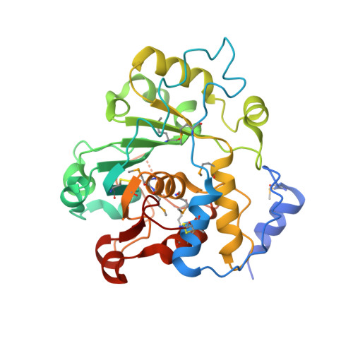
Glycan structures on glycoproteins and glycolipids play critical roles in biological recognition, targeting, and modulation of functions in animal systems. Many classes of glycan structures are capped with terminal sialic acid residues, which contribute to biological functions by either forming or masking glycan recognition sites on the cell surface or secreted glycoconjugates. Sialylated glycans are synthesized in mammals by a single conserved family of sialyltransferases that have diverse linkage and acceptor specificities. We examined the enzymatic basis for glycan sialylation in animal systems by determining the crystal structures of rat ST6GAL1, an enzyme that creates terminal α2,6-sialic acid linkages on complex-type N-glycans, at 2.4 Å resolution. Crystals were obtained from enzyme preparations generated in mammalian cells. The resulting structure revealed an overall protein fold broadly resembling the previously determined structure of pig ST3GAL1, including a CMP-sialic acid-binding site assembled from conserved sialylmotif sequence elements. Significant differences in structure and disulfide bonding patterns were found outside the sialylmotif sequences, including differences in residues predicted to interact with the glycan acceptor. Computational substrate docking and molecular dynamics simulations were performed to predict and evaluate the CMP-sialic acid donor and glycan acceptor interactions, and the results were compared with kinetic analysis of active site mutants. Comparisons of the structure with pig ST3GAL1 and a bacterial sialyltransferase revealed a similar positioning of donor, acceptor, and catalytic residues that provide a common structural framework for catalysis by the mammalian and bacterial sialyltransferases.
Organizational Affiliation:
From the Complex Carbohydrate Research Center, University of Georgia, Athens, Georgia 30602.