4MMF

Crystal structure of LeuBAT (delta5 mutant) in complex with mazindol


Experimental Data Snapshot

  • Method: X-RAY DIFFRACTION
  • Resolution: 2.70 Å
  • R-Value Free: 0.236 
  • R-Value Work: 0.199 
  • R-Value Observed: 0.201 

wwPDB Validation   3D Report Full Report


Ligand Structure Quality Assessment 


This is version 1.3 of the entry. See complete history


Literature

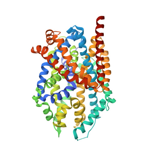
Structural basis for action by diverse antidepressants on biogenic amine transporters.

Wang, H.Goehring, A.Wang, K.H.Penmatsa, A.Ressler, R.Gouaux, E.

(2013) Nature 503: 141-145

  • DOI: https://doi.org/10.1038/nature12648
  • Primary Citation of Related Structures:  
    4MM4, 4MM5, 4MM6, 4MM7, 4MM8, 4MM9, 4MMA, 4MMB, 4MMC, 4MMD, 4MME, 4MMF

  • PubMed Abstract: 

    The biogenic amine transporters (BATs) regulate endogenous neurotransmitter concentrations and are targets for a broad range of therapeutic agents including selective serotonin reuptake inhibitors (SSRIs), serotonin-noradrenaline reuptake inhibitors (SNRIs) and tricyclic antidepressants (TCAs). Because eukaryotic BATs are recalcitrant to crystallographic analysis, our understanding of the mechanism of these inhibitors and antidepressants is limited. LeuT is a bacterial homologue of BATs and has proven to be a valuable paradigm for understanding relationships between their structure and function. However, because only approximately 25% of the amino acid sequence of LeuT is in common with that of BATs, and as LeuT is a promiscuous amino acid transporter, it does not recapitulate the pharmacological properties of BATs. Indeed, SSRIs and TCAs bind in the extracellular vestibule of LeuT and act as non-competitive inhibitors of transport. By contrast, multiple studies demonstrate that both TCAs and SSRIs are competitive inhibitors for eukaryotic BATs and bind to the primary binding pocket. Here we engineered LeuT to harbour human BAT-like pharmacology by mutating key residues around the primary binding pocket. The final LeuBAT mutant binds the SSRI sertraline with a binding constant of 18 nM and displays high-affinity binding to a range of SSRIs, SNRIs and a TCA. We determined 12 crystal structures of LeuBAT in complex with four classes of antidepressants. The chemically diverse inhibitors have a remarkably similar mode of binding in which they straddle transmembrane helix (TM) 3, wedge between TM3/TM8 and TM1/TM6, and lock the transporter in a sodium- and chloride-bound outward-facing open conformation. Together, these studies define common and simple principles for the action of SSRIs, SNRIs and TCAs on BATs.


  • Organizational Affiliation

    Vollum Institute, Oregon Health & Science University, 3181 SW Sam Jackson Park Road, Portland, Oregon 97239, USA.


Macromolecules
Find similar proteins by:  (by identity cutoff)  |  3D Structure
Entity ID: 1
MoleculeChains Sequence LengthOrganismDetailsImage
Transporter
A, B
518Aquifex aeolicus VF5Mutation(s): 5 
Gene Names: snfaq_2077
Membrane Entity: Yes 
UniProt
Find proteins for O67854 (Aquifex aeolicus (strain VF5))
Explore O67854 
Go to UniProtKB:  O67854
Entity Groups  
Sequence Clusters30% Identity50% Identity70% Identity90% Identity95% Identity100% Identity
UniProt GroupO67854
Sequence Annotations
Expand
  • Reference Sequence
Small Molecules
Ligands 3 Unique
IDChains Name / Formula / InChI Key2D Diagram3D Interactions
BOG
Query on BOG

Download Ideal Coordinates CCD File 
F [auth A],
G [auth A],
K [auth B],
L [auth B],
M [auth B]
octyl beta-D-glucopyranoside
C14 H28 O6
HEGSGKPQLMEBJL-RKQHYHRCSA-N
29Q
Query on 29Q

Download Ideal Coordinates CCD File 
E [auth A],
J [auth B]
(5R)-5-(4-chlorophenyl)-2,5-dihydro-3H-imidazo[2,1-a]isoindol-5-ol
C16 H13 Cl N2 O
ZPXSCAKFGYXMGA-MRXNPFEDSA-N
NA
Query on NA

Download Ideal Coordinates CCD File 
C [auth A],
D [auth A],
H [auth B],
I [auth B]
SODIUM ION
Na
FKNQFGJONOIPTF-UHFFFAOYSA-N
Binding Affinity Annotations 
IDSourceBinding Affinity
29Q PDBBind:  4MMF Kd: 295 (nM) from 1 assay(s)
Experimental Data & Validation

Experimental Data

  • Method: X-RAY DIFFRACTION
  • Resolution: 2.70 Å
  • R-Value Free: 0.236 
  • R-Value Work: 0.199 
  • R-Value Observed: 0.201 
  • Space Group: P 1 21 1
Unit Cell:
Length ( Å )Angle ( ˚ )
a = 84.227α = 90
b = 92.137β = 93.54
c = 87.827γ = 90
Software Package:
Software NamePurpose
ADSCdata collection
PHASERphasing
PHENIXrefinement
HKL-2000data reduction
HKL-2000data scaling

Structure Validation

View Full Validation Report


Ligand Structure Quality Assessment 


Entry History 

Deposition Data

Revision History  (Full details and data files)

  • Version 1.0: 2013-10-16
    Type: Initial release
  • Version 1.1: 2013-11-20
    Changes: Database references
  • Version 1.2: 2020-07-29
    Type: Remediation
    Reason: Carbohydrate remediation
    Changes: Data collection, Database references, Derived calculations, Structure summary
  • Version 1.3: 2024-02-28
    Changes: Data collection, Database references, Structure summary