The Multidrug Resistance IncA/C Transferable Plasmid Encodes a Novel Domain-swapped Dimeric Protein-disulfide Isomerase.
Premkumar, L., Kurth, F., Neyer, S., Schembri, M.A., Martin, J.L.(2014) J Biol Chem 289: 2563-2576
- PubMed: 24311786
- DOI: https://doi.org/10.1074/jbc.M113.516898
- Primary Citation of Related Structures:
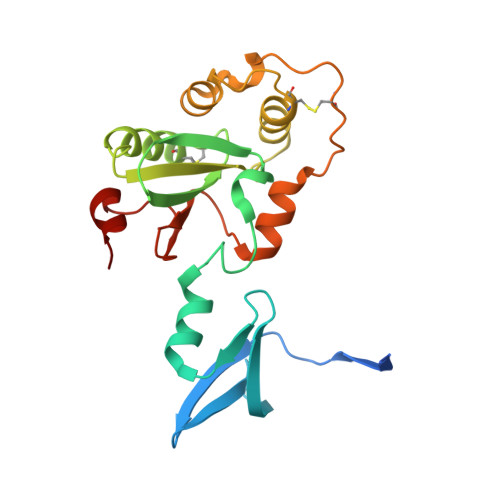
4ML1, 4ML6, 4MLY - PubMed Abstract:
The multidrug resistance-encoding IncA/C conjugative plasmids disseminate antibiotic resistance genes among clinically relevant enteric bacteria. A plasmid-encoded disulfide isomerase is associated with conjugation. Sequence analysis of several IncA/C plasmids and IncA/C-related integrative and conjugative elements (ICE) from commensal and pathogenic bacteria identified a conserved DsbC/DsbG homolog (DsbP). The crystal structure of DsbP reveals an N-terminal domain, a linker region, and a C-terminal catalytic domain. A DsbP homodimer is formed through domain swapping of two DsbP N-terminal domains. The catalytic domain incorporates a thioredoxin-fold with characteristic CXXC and cis-Pro motifs. Overall, the structure and redox properties of DsbP diverge from the Escherichia coli DsbC and DsbG disulfide isomerases. Specifically, the V-shaped dimer of DsbP is inverted compared with EcDsbC and EcDsbG. In addition, the redox potential of DsbP (-161 mV) is more reducing than EcDsbC (-130 mV) and EcDsbG (-126 mV). Other catalytic properties of DsbP more closely resemble those of EcDsbG than EcDsbC. These catalytic differences are in part a consequence of the unusual active site motif of DsbP (CAVC); substitution to the EcDsbC-like (CGYC) motif converts the catalytic properties to those of EcDsbC. Structural comparison of the 12 independent subunit structures of DsbP that we determined revealed that conformational changes in the linker region contribute to mobility of the catalytic domain, providing mechanistic insight into DsbP function. In summary, our data reveal that the conserved plasmid-encoded DsbP protein is a bona fide disulfide isomerase and suggest that a dedicated oxidative folding enzyme is important for conjugative plasmid transfer.
Organizational Affiliation:
From the Institute for Molecular Bioscience, Division of Chemistry and Structural Biology and.