


Crystal structure of extracellular ligand-binding receptor from Verminephrobacter eiseniae ef01-2
Wu, R., Endres, M., Joachimiak, A., Midwest Center for Structural GenomicsTo be published.
Experimental Data Snapshot
wwPDB Validation 3D Report Full Report

Entity ID: 1 | |||||
|---|---|---|---|---|---|
| Molecule | Chains | Sequence Length | Organism | Details | Image |
| Extracellular ligand-binding receptor | 357 | Verminephrobacter eiseniae EF01-2 | Mutation(s): 0 Gene Names: Veis_2660 | 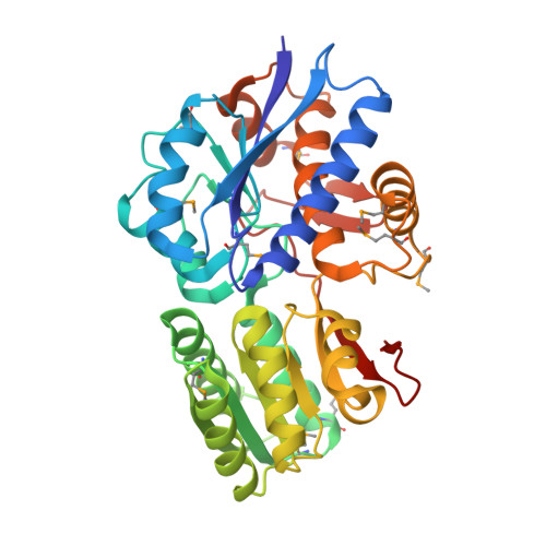 | |
UniProt | |||||
Find proteins for A1WL96 (Verminephrobacter eiseniae (strain EF01-2)) Explore A1WL96 Go to UniProtKB: A1WL96 | |||||
Entity Groups | |||||
| Sequence Clusters | 30% Identity50% Identity70% Identity90% Identity95% Identity100% Identity | ||||
| UniProt Group | A1WL96 | ||||
Sequence AnnotationsExpand | |||||
| |||||
| Ligands 1 Unique | |||||
|---|---|---|---|---|---|
| ID | Chains | Name / Formula / InChI Key | 2D Diagram | 3D Interactions | |
| GOL Query on GOL | B [auth A], C [auth A], D [auth A], E [auth A] | GLYCEROL C3 H8 O3 PEDCQBHIVMGVHV-UHFFFAOYSA-N |  | ||
| Modified Residues 1 Unique | |||||
|---|---|---|---|---|---|
| ID | Chains | Type | Formula | 2D Diagram | Parent |
| MSE Query on MSE | A | L-PEPTIDE LINKING | C5 H11 N O2 Se |  | MET |
| Length ( Å ) | Angle ( ˚ ) |
|---|---|
| a = 36.076 | α = 90 |
| b = 95.81 | β = 111.56 |
| c = 50.3 | γ = 90 |
| Software Name | Purpose |
|---|---|
| HKL-3000 | data collection |
| MLPHARE | phasing |
| PHENIX | refinement |
| HKL-3000 | data reduction |
| HKL-3000 | data scaling |
RCSB PDB (citation) is hosted by
RCSB PDB is a member of the

