


The crystal structure of a possible an iron-binding (periplasmic solute-binding) protein from Staphylococcus epidermidis ATCC 12228.
Tan, K., Li, H., Clancy, S., Joachimiak, A.To be published.
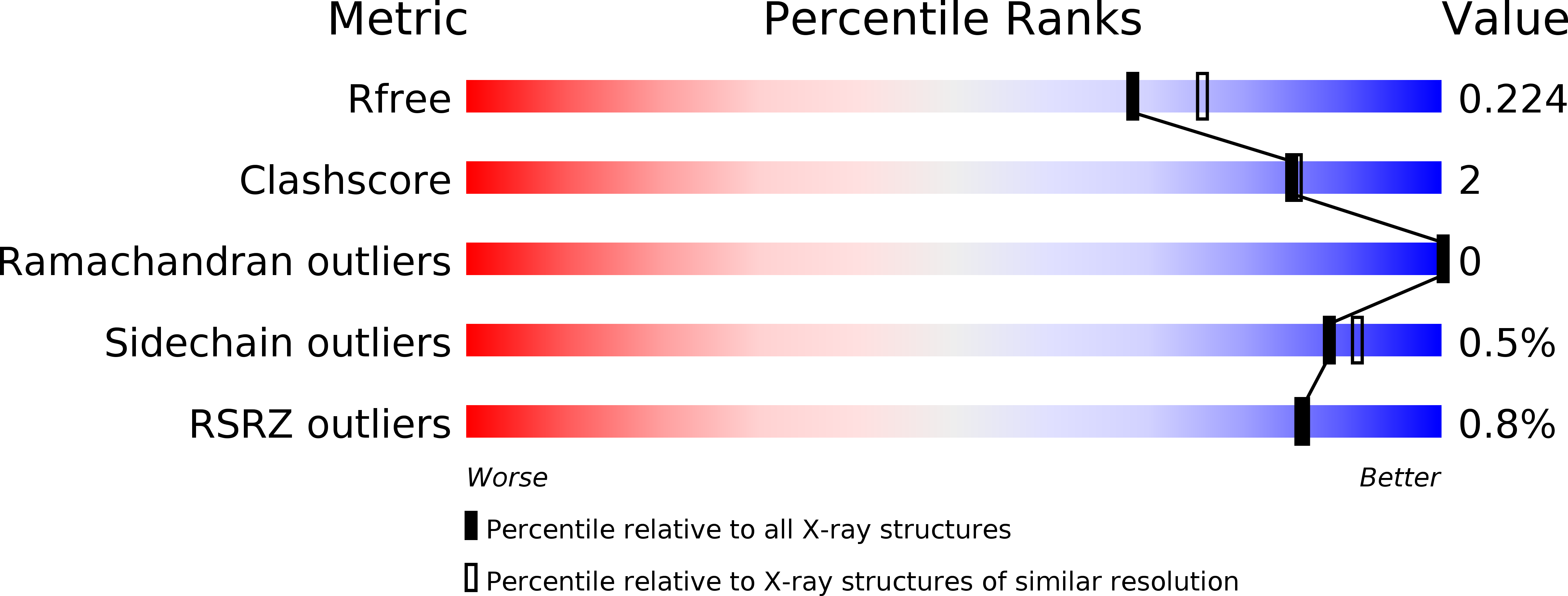
Experimental Data Snapshot
wwPDB Validation 3D Report Full Report
Entity ID: 1 | |||||
|---|---|---|---|---|---|
| Molecule | Chains | Sequence Length | Organism | Details | Image |
| Iron-binding protein | 278 | Staphylococcus epidermidis ATCC 12228 | Mutation(s): 0 Gene Names: SE_0383 |  | |
UniProt | |||||
Find proteins for A0A0H2VHB1 (Staphylococcus epidermidis (strain ATCC 12228 / FDA PCI 1200)) Explore A0A0H2VHB1 Go to UniProtKB: A0A0H2VHB1 | |||||
Entity Groups | |||||
| Sequence Clusters | 30% Identity50% Identity70% Identity90% Identity95% Identity100% Identity | ||||
| UniProt Group | A0A0H2VHB1 | ||||
Sequence AnnotationsExpand | |||||
| |||||
| Modified Residues 1 Unique | |||||
|---|---|---|---|---|---|
| ID | Chains | Type | Formula | 2D Diagram | Parent |
| MSE Query on MSE | A | L-PEPTIDE LINKING | C5 H11 N O2 Se |  | MET |
| Length ( Å ) | Angle ( ˚ ) |
|---|---|
| a = 154.052 | α = 90 |
| b = 48.173 | β = 102.74 |
| c = 44.916 | γ = 90 |
| Software Name | Purpose |
|---|---|
| SBC-Collect | data collection |
| SHELXD | phasing |
| MLPHARE | phasing |
| DM | model building |
| ARP | model building |
| WARP | model building |
| HKL-3000 | phasing |
| PHENIX | refinement |
| HKL-3000 | data reduction |
| HKL-3000 | data scaling |
| DM | phasing |
RCSB PDB (citation) is hosted by
RCSB PDB is a member of the

