4M7K

Crystal structure of anti-tissue factor antibody 10H10


Experimental Data Snapshot

  • Method: X-RAY DIFFRACTION
  • Resolution: 1.90 Å
  • R-Value Free: 0.243 
  • R-Value Work: 0.205 
  • R-Value Observed: 0.206 

wwPDB Validation   3D Report Full Report


This is version 2.1 of the entry. See complete history


Literature

Antibody modeling assessment II. Structures and models.

Teplyakov, A.Luo, J.Obmolova, G.Malia, T.J.Sweet, R.Stanfield, R.L.Kodangattil, S.Almagro, J.C.Gilliland, G.L.

(2014) Proteins 82: 1563-1582

  • DOI: https://doi.org/10.1002/prot.24554
  • Primary Citation of Related Structures:  
    4KMT, 4KQ4, 4M6M, 4M6O, 4M7K, 4MAU

  • PubMed Abstract: 

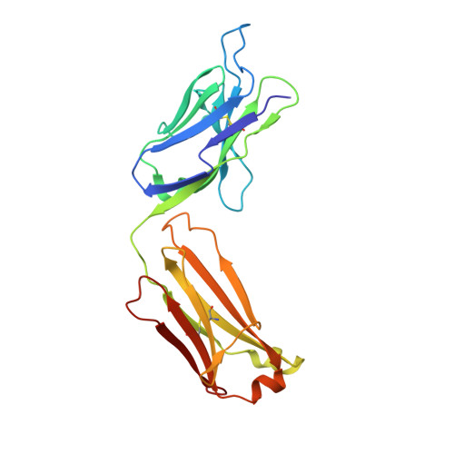
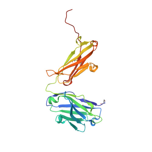
    To assess the state-of-the-art in antibody structure modeling, a blinded study was conducted. Eleven unpublished Fab crystal structures were used as a benchmark to compare Fv models generated by seven structure prediction methodologies. In the first round, each participant submitted three non-ranked complete Fv models for each target. In the second round, CDR-H3 modeling was performed in the context of the correct environment provided by the crystal structures with CDR-H3 removed. In this report we describe the reference structures and present our assessment of the models. Some of the essential sources of errors in the predictions were traced to the selection of the structure template, both in terms of the CDR canonical structures and VL/VH packing. On top of this, the errors present in the Protein Data Bank structures were sometimes propagated in the current models, which emphasized the need for the curated structural database devoid of errors. Modeling non-canonical structures, including CDR-H3, remains the biggest challenge for antibody structure prediction.


  • Organizational Affiliation

    Janssen Research & Development, LLC, 1400 McKean Road, Spring House, Pennsylvania, 19477.


Macromolecules
Find similar proteins by:  (by identity cutoff)  |  3D Structure
Entity ID: 1
MoleculeChains Sequence LengthOrganismDetailsImage
10H10 light chainA [auth L]220Mus musculusHomo sapiens
This entity is chimeric
Mutation(s): 0 
Entity Groups  
Sequence Clusters30% Identity50% Identity70% Identity90% Identity95% Identity100% Identity
Sequence Annotations
Expand
  • Reference Sequence
Find similar proteins by:  (by identity cutoff)  |  3D Structure
Entity ID: 2
MoleculeChains Sequence LengthOrganismDetailsImage
10H10 heavy chainB [auth H]229Mus musculusHomo sapiens
This entity is chimeric
Mutation(s): 0 
Entity Groups  
Sequence Clusters30% Identity50% Identity70% Identity90% Identity95% Identity100% Identity
Sequence Annotations
Expand
  • Reference Sequence
Small Molecules
Modified Residues  1 Unique
IDChains TypeFormula2D DiagramParent
PCA
Query on PCA
B [auth H]L-PEPTIDE LINKINGC5 H7 N O3GLN
Experimental Data & Validation

Experimental Data

  • Method: X-RAY DIFFRACTION
  • Resolution: 1.90 Å
  • R-Value Free: 0.243 
  • R-Value Work: 0.205 
  • R-Value Observed: 0.206 
  • Space Group: C 2 2 21
Unit Cell:
Length ( Å )Angle ( ˚ )
a = 81.77α = 90
b = 135.51β = 90
c = 88.25γ = 90
Software Package:
Software NamePurpose
StructureStudiodata collection
AMoREphasing
REFMACrefinement
XDSdata reduction
XDSdata scaling

Structure Validation

View Full Validation Report


Entry History 

Deposition Data

Revision History  (Full details and data files)

  • Version 1.0: 2014-03-26
    Type: Initial release
  • Version 1.1: 2014-04-02
    Changes: Database references
  • Version 1.2: 2014-08-06
    Changes: Database references
  • Version 2.0: 2019-12-25
    Changes: Derived calculations, Polymer sequence
  • Version 2.1: 2023-09-20
    Changes: Data collection, Database references, Derived calculations, Refinement description