4M61

Crystal structure of unliganded anti-DNA Fab A52


Experimental Data Snapshot

  • Method: X-RAY DIFFRACTION
  • Resolution: 1.62 Å
  • R-Value Free: 0.232 
  • R-Value Work: 0.192 
  • R-Value Observed: 0.196 

wwPDB Validation   3D Report Full Report


This is version 1.3 of the entry. See complete history


Literature

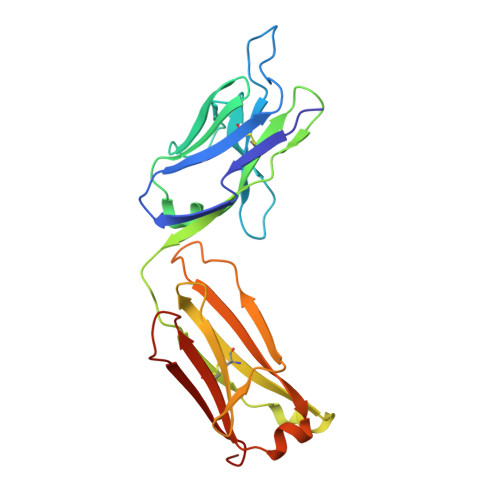
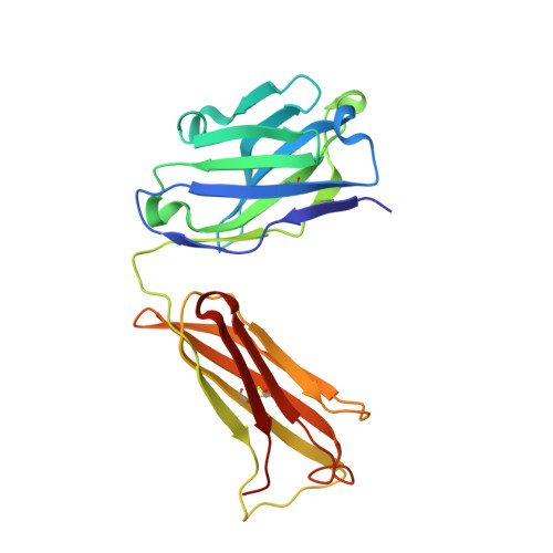
Crystal structure determination of anti-DNA Fab A52.

Stanfield, R.L.Eilat, D.

(2014) Proteins 82: 1674-1678

  • DOI: https://doi.org/10.1002/prot.24514
  • Primary Citation of Related Structures:  
    4M61

  • PubMed Abstract: 

    A52 is a murine monoclonal antibody isolated from autoimmune New Zealand Black/New Zealand White F1 mice that recognizes single and double stranded DNA. This mouse strain spontaneously develops systemic lupus erythematosus-like symptoms and has served as a model for that disease for many years. The 1.62 Å crystal structure of the A52 Fab fragment reveals an H3 complementarity determining region with four closely spaced arginine residues, creating a positively charged surface to accommodate bound DNA.


  • Organizational Affiliation

    The Department of Integrative Structural and Computational Biology, The Scripps Research Institute, La Jolla, California, 92037.


Macromolecules
Find similar proteins by:  (by identity cutoff)  |  3D Structure
Entity ID: 1
MoleculeChains Sequence LengthOrganismDetailsImage
Fab A52 light chain
A, C
219Mus musculusMutation(s): 0 
Entity Groups  
Sequence Clusters30% Identity50% Identity70% Identity90% Identity95% Identity100% Identity
Sequence Annotations
Expand
  • Reference Sequence
Find similar proteins by:  (by identity cutoff)  |  3D Structure
Entity ID: 2
MoleculeChains Sequence LengthOrganismDetailsImage
Fab A52 heavy chain
B, D
220Mus musculusMutation(s): 0 
Entity Groups  
Sequence Clusters30% Identity50% Identity70% Identity90% Identity95% Identity100% Identity
Sequence Annotations
Expand
  • Reference Sequence
Small Molecules
Ligands 1 Unique
IDChains Name / Formula / InChI Key2D Diagram3D Interactions
SO4
Query on SO4

Download Ideal Coordinates CCD File 
E [auth A]
F [auth A]
G [auth A]
H [auth A]
I [auth B]
E [auth A],
F [auth A],
G [auth A],
H [auth A],
I [auth B],
J [auth B],
K [auth B],
L [auth B],
M [auth B],
N [auth B],
O [auth C],
P [auth C],
Q [auth C],
R [auth C],
S [auth C],
T [auth D],
U [auth D],
V [auth D],
W [auth D]
SULFATE ION
O4 S
QAOWNCQODCNURD-UHFFFAOYSA-L
Experimental Data & Validation

Experimental Data

  • Method: X-RAY DIFFRACTION
  • Resolution: 1.62 Å
  • R-Value Free: 0.232 
  • R-Value Work: 0.192 
  • R-Value Observed: 0.196 
  • Space Group: P 1 21 1
Unit Cell:
Length ( Å )Angle ( ˚ )
a = 52.784α = 90
b = 61.38β = 96.51
c = 121.223γ = 90
Software Package:
Software NamePurpose
EPMRphasing
PHENIXrefinement
DENZOdata reduction
SCALEPACKdata scaling

Structure Validation

View Full Validation Report


Entry History 

Deposition Data

Revision History  (Full details and data files)

  • Version 1.0: 2013-08-28
    Type: Initial release
  • Version 1.1: 2014-02-26
    Changes: Database references
  • Version 1.2: 2014-08-06
    Changes: Database references
  • Version 1.3: 2023-09-20
    Changes: Data collection, Database references, Derived calculations, Refinement description