4LP9

Endothiapepsin complexed with Phe-reduced-Tyr peptide.


Experimental Data Snapshot

  • Method: X-RAY DIFFRACTION
  • Resolution: 1.35 Å
  • R-Value Free: 0.165 
  • R-Value Work: 0.123 
  • R-Value Observed: 0.125 

wwPDB Validation   3D Report Full Report


This is version 2.0 of the entry. See complete history


Literature

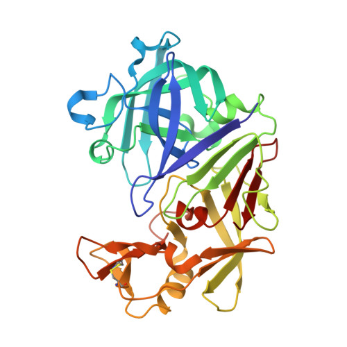
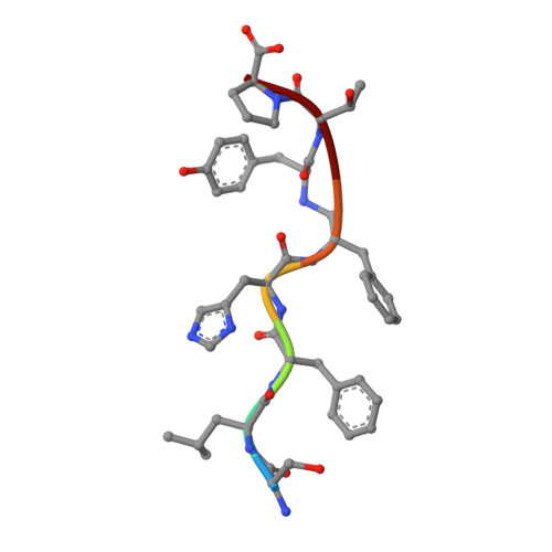
The structure of endothiapepsin complexed with a Phe-Tyr reduced-bond inhibitor at 1.35 angstrom resolution.

Guo, J.Cooper, J.B.Wood, S.P.

(2014) Acta Crystallogr D Biol Crystallogr 70: 30-33

  • DOI: https://doi.org/10.1107/S2053230X13032974
  • Primary Citation of Related Structures:  
    4LP9

  • PubMed Abstract: 

    Endothiapepsin is a typical member of the aspartic proteinase family. The catalytic mechanism of this family is attributed to two conserved catalytic aspartate residues, which coordinate the hydrolysis of a peptide bond. An oligopeptide inhibitor (IC50 = 0.62 µM) based on a reduced-bond transition-state inhibitor of mucorpepsin was co-crystallized with endothiapepsin and the crystal structure of the enzyme-inhibitor complex was determined at 1.35 Å resolution. A total of 12 hydrogen bonds between the inhibitor and the active-site residues were identified. The resulting structure demonstrates a number of novel subsite interactions in the active-site cleft.


  • Organizational Affiliation

    Laboratory of Protein Crystallography, Centre for Amyloidosis and Acute Phase Proteins, UCL Division of Medicine (Royal Free Campus), Rowland Hill Street, London NW3 2PF, England.


Macromolecules
Find similar proteins by:  (by identity cutoff)  |  3D Structure
Entity ID: 1
MoleculeChains Sequence LengthOrganismDetailsImage
Endothiapepsin330Cryphonectria parasiticaMutation(s): 0 
EC: 3.4.23.22
UniProt
Find proteins for P11838 (Cryphonectria parasitica)
Explore P11838 
Go to UniProtKB:  P11838
Entity Groups  
Sequence Clusters30% Identity50% Identity70% Identity90% Identity95% Identity100% Identity
UniProt GroupP11838
Sequence Annotations
Expand
  • Reference Sequence

Find similar proteins by:  Sequence   |   3D Structure  

Entity ID: 2
MoleculeChains Sequence LengthOrganismDetailsImage
Ser-Leu-Phe-His-Phenylalanyl-reduced-peptide-bond-Tyrosyl-Thr-ProB [auth I]7N/AMutation(s): 0 
Sequence Annotations
Expand
  • Reference Sequence
Biologically Interesting Molecules (External Reference) 1 Unique
Experimental Data & Validation

Experimental Data

  • Method: X-RAY DIFFRACTION
  • Resolution: 1.35 Å
  • R-Value Free: 0.165 
  • R-Value Work: 0.123 
  • R-Value Observed: 0.125 
  • Space Group: P 1 21 1
Unit Cell:
Length ( Å )Angle ( ˚ )
a = 52.62α = 90
b = 72.787β = 108.54
c = 45.113γ = 90
Software Package:
Software NamePurpose
GDAdata collection
FFTmodel building
REFMACrefinement
XDSdata reduction
SCALAdata scaling
FFTphasing

Structure Validation

View Full Validation Report


Entry History 

Deposition Data

Revision History  (Full details and data files)

  • Version 1.0: 2014-01-15
    Type: Initial release
  • Version 1.1: 2014-02-19
    Changes: Database references
  • Version 1.2: 2023-09-20
    Changes: Data collection, Database references, Derived calculations, Refinement description
  • Version 2.0: 2023-11-15
    Changes: Atomic model, Data collection, Derived calculations