4LLF

Crystal structure of Cucumber Necrosis Virus


Experimental Data Snapshot

  • Method: X-RAY DIFFRACTION
  • Resolution: 2.89 Å
  • R-Value Free: 0.242 
  • R-Value Work: 0.213 
  • R-Value Observed: 0.214 

wwPDB Validation   3D Report Full Report


This is version 1.1 of the entry. See complete history


Literature

Atomic structure of cucumber necrosis virus and the role of the capsid in vector transmission.

Li, M.Kakani, K.Katpally, U.Johnson, S.Rochon, D.Smith, T.J.

(2013) J Virol 87: 12166-12175

  • DOI: https://doi.org/10.1128/JVI.01965-13
  • Primary Citation of Related Structures:  
    4LLF

  • PubMed Abstract: 

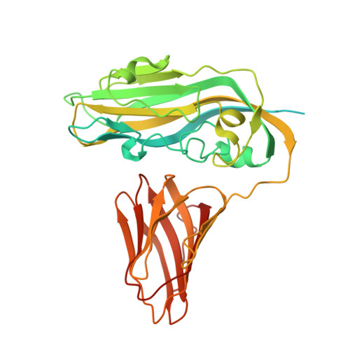
    Cucumber Necrosis Virus (CNV) is a member of the genus Tombusvirus and has a monopartite positive-sense RNA genome packaged in a T=3 icosahedral particle. CNV is transmitted in nature via zoospores of the fungus Olpidium bornovanus. CNV undergoes a conformational change upon binding to the zoospore that is required for transmission, and specific polysaccharides on the zoospore surface have been implicated in binding. To better understand this transmission process, we have determined the atomic structure of CNV. As expected, being a member of the Tombusvirus genus, the core structure of CNV is highly similar to that of Tomato bushy stunt virus (TBSV), with major differences lying on the exposed loops. Also, as was seen with TBSV, CNV appears to have a calcium binding site between the subunits around the quasi-3-fold axes. However, unlike TBSV, there appears to be a novel zinc binding site within the β annulus formed by the N termini of the three C subunits at the icosahedral 3-fold axes. Two of the mutations causing defective transmission map immediately around this zinc binding site. The other mutations causing defective transmission and particle formation are mapped onto the CNV structure, and it is likely that a number of the mutations affect zoospore transmission by affecting conformational transitions rather than directly affecting receptor binding.


  • Organizational Affiliation

    Donald Danforth Plant Science Center, St. Louis, Missouri, USA.


Macromolecules
Find similar proteins by:  (by identity cutoff)  |  3D Structure
Entity ID: 1
MoleculeChains Sequence LengthOrganismDetailsImage
Capsid protein380Cucumber necrosis virusMutation(s): 0 
UniProt
Find proteins for P15183 (Cucumber necrosis virus)
Explore P15183 
Go to UniProtKB:  P15183
Entity Groups  
Sequence Clusters30% Identity50% Identity70% Identity90% Identity95% Identity100% Identity
UniProt GroupP15183
Sequence Annotations
Expand
  • Reference Sequence
Small Molecules
Ligands 2 Unique
IDChains Name / Formula / InChI Key2D Diagram3D Interactions
ZN
Query on ZN

Download Ideal Coordinates CCD File 
AA [auth J],
DA [auth M],
IA [auth P],
R [auth D],
W [auth G]
ZINC ION
Zn
PTFCDOFLOPIGGS-UHFFFAOYSA-N
CA
Query on CA

Download Ideal Coordinates CCD File 
BA [auth K]
CA [auth K]
EA [auth M]
FA [auth N]
GA [auth O]
BA [auth K],
CA [auth K],
EA [auth M],
FA [auth N],
GA [auth O],
HA [auth O],
P [auth B],
Q [auth B],
S [auth D],
T [auth E],
U [auth E],
V [auth F],
X [auth H],
Y [auth I],
Z [auth I]
CALCIUM ION
Ca
BHPQYMZQTOCNFJ-UHFFFAOYSA-N
Experimental Data & Validation

Experimental Data

  • Method: X-RAY DIFFRACTION
  • Resolution: 2.89 Å
  • R-Value Free: 0.242 
  • R-Value Work: 0.213 
  • R-Value Observed: 0.214 
  • Space Group: I 2 3
Unit Cell:
Length ( Å )Angle ( ˚ )
a = 384α = 90
b = 384β = 90
c = 384γ = 90
Software Package:
Software NamePurpose
PROTEUM PLUSdata collection
PHENIXmodel building
PHENIXrefinement
SAINTdata reduction
SHELXdata scaling
PHENIXphasing

Structure Validation

View Full Validation Report


Entry History 

Deposition Data

  • Released Date: 2013-10-30 
  • Deposition Author(s): Smith, T.

Revision History  (Full details and data files)

  • Version 1.0: 2013-10-30
    Type: Initial release
  • Version 1.1: 2023-09-20
    Changes: Data collection, Database references, Derived calculations, Refinement description