4LEX

Unliganded crystal structure of mAb7


Experimental Data Snapshot

  • Method: X-RAY DIFFRACTION
  • Resolution: 2.02 Å
  • R-Value Free: 0.218 
  • R-Value Work: 0.185 
  • R-Value Observed: 0.187 

wwPDB Validation   3D Report Full Report


This is version 1.3 of the entry. See complete history


Literature

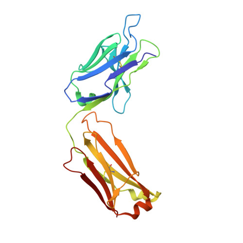
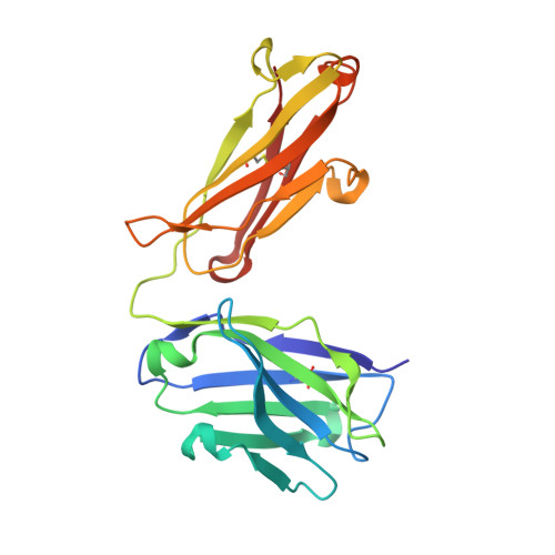
Inhibitory mechanism of an allosteric antibody targeting the glucagon receptor.

Mukund, S.Shang, Y.Clarke, H.J.Madjidi, A.Corn, J.E.Kates, L.Kolumam, G.Chiang, V.Luis, E.Murray, J.Zhang, Y.Hotzel, I.Koth, C.M.Allan, B.B.

(2013) J Biol Chem 288: 36168-36178

  • DOI: https://doi.org/10.1074/jbc.M113.496984
  • Primary Citation of Related Structures:  
    4LEX, 4LF3

  • PubMed Abstract: 

    Elevated glucagon levels and increased hepatic glucagon receptor (GCGR) signaling contribute to hyperglycemia in type 2 diabetes. We have identified a monoclonal antibody that inhibits GCGR, a class B G-protein coupled receptor (GPCR), through a unique allosteric mechanism. Receptor inhibition is mediated by the binding of this antibody to two distinct sites that lie outside of the glucagon binding cleft. One site consists of a patch of residues that are surface-exposed on the face of the extracellular domain (ECD) opposite the ligand-binding cleft, whereas the second binding site consists of residues in the αA helix of the ECD. A docking model suggests that the antibody does not occlude the ligand-binding cleft. We solved the crystal structure of GCGR ECD containing a naturally occurring G40S mutation and found a shift in the register of the αA helix that prevents antibody binding. We also found that alterations in the αA helix impact the normal function of GCGR. We present a model for the allosteric inhibition of GCGR by a monoclonal antibody that may form the basis for the development of allosteric modulators for the treatment of diabetes and other class B GPCR-related diseases.


  • Organizational Affiliation

    From the Departments of Structural Biology.


Macromolecules
Find similar proteins by:  (by identity cutoff)  |  3D Structure
Entity ID: 1
MoleculeChains Sequence LengthOrganismDetailsImage
FAB HEAVY CHAINA [auth L]217Mus musculusMutation(s): 0 
Entity Groups  
Sequence Clusters30% Identity50% Identity70% Identity90% Identity95% Identity100% Identity
Sequence Annotations
Expand
  • Reference Sequence
Find similar proteins by:  (by identity cutoff)  |  3D Structure
Entity ID: 2
MoleculeChains Sequence LengthOrganismDetailsImage
FAB LIGHT CHAINB [auth H]217Mus musculusMutation(s): 0 
Entity Groups  
Sequence Clusters30% Identity50% Identity70% Identity90% Identity95% Identity100% Identity
Sequence Annotations
Expand
  • Reference Sequence
Experimental Data & Validation

Experimental Data

  • Method: X-RAY DIFFRACTION
  • Resolution: 2.02 Å
  • R-Value Free: 0.218 
  • R-Value Work: 0.185 
  • R-Value Observed: 0.187 
  • Space Group: P 21 2 21
Unit Cell:
Length ( Å )Angle ( ˚ )
a = 59.069α = 90
b = 84.184β = 90
c = 88.959γ = 90
Software Package:
Software NamePurpose
DENZOdata reduction
SCALEPACKdata scaling
PHASERphasing
PHENIXrefinement
PDB_EXTRACTdata extraction
ADSCdata collection

Structure Validation

View Full Validation Report


Entry History 

Deposition Data

Revision History  (Full details and data files)

  • Version 1.0: 2013-11-13
    Type: Initial release
  • Version 1.1: 2013-11-27
    Changes: Database references
  • Version 1.2: 2014-01-01
    Changes: Database references
  • Version 1.3: 2023-09-20
    Changes: Data collection, Database references, Refinement description