4L4S

Structural characterisation of the NADH binary complex of human lactate dehydrogenase M isozyme


Experimental Data Snapshot

  • Method: X-RAY DIFFRACTION
  • Resolution: 2.90 Å
  • R-Value Free: 0.246 
  • R-Value Work: 0.185 
  • R-Value Observed: 0.188 

wwPDB Validation   3D Report Full Report


Ligand Structure Quality Assessment 


This is version 1.3 of the entry. See complete history


Literature

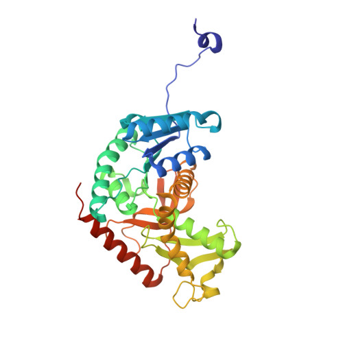
Structural characterization of the apo form and NADH binary complex of human lactate dehydrogenase.

Dempster, S.Harper, S.Moses, J.E.Dreveny, I.

(2014) Acta Crystallogr D Biol Crystallogr 70: 1484-1490

  • DOI: https://doi.org/10.1107/S1399004714005422
  • Primary Citation of Related Structures:  
    4L4R, 4L4S

  • PubMed Abstract: 

    Lactate dehydrogenase A (LDH-A) is a key enzyme in anaerobic respiration that is predominantly found in skeletal muscle and catalyses the reversible conversion of pyruvate to lactate in the presence of NADH. LDH-A is overexpressed in many tumours and has therefore emerged as an attractive target for anticancer drug discovery. Crystal structures of human LDH-A in the presence of inhibitors have been described, but currently no structures of the apo or binary NADH-bound forms are available for any mammalian LDH-A. Here, the apo structure of human LDH-A was solved at a resolution of 2.1 Å in space group P4122. The active-site loop adopts an open conformation and the packing and crystallization conditions suggest that the crystal form is suitable for soaking experiments. The soaking potential was assessed with the cofactor NADH, which yielded a ligand-bound crystal structure in the absence of any inhibitors. The structures show that NADH binding induces small conformational changes in the active-site loop and an adjacent helix. A comparison with other eukaryotic apo LDH structures reveals the conservation of intra-loop interactions. The structures provide novel insight into cofactor binding and provide the foundation for soaking experiments with fragments and inhibitors.


  • Organizational Affiliation

    School of Chemistry, University of Nottingham, Nottingham NG7 2RD, England.


Macromolecules
Find similar proteins by:  (by identity cutoff)  |  3D Structure
Entity ID: 1
MoleculeChains Sequence LengthOrganismDetailsImage
L-lactate dehydrogenase A chainA [auth H],
B [auth A]
339Homo sapiensMutation(s): 0 
Gene Names: LDHAPIG19
EC: 1.1.1.27
UniProt & NIH Common Fund Data Resources
Find proteins for P00338 (Homo sapiens)
Explore P00338 
Go to UniProtKB:  P00338
PHAROS:  P00338
GTEx:  ENSG00000134333 
Entity Groups  
Sequence Clusters30% Identity50% Identity70% Identity90% Identity95% Identity100% Identity
UniProt GroupP00338
Sequence Annotations
Expand
  • Reference Sequence
Small Molecules
Ligands 1 Unique
IDChains Name / Formula / InChI Key2D Diagram3D Interactions
NAI
Query on NAI

Download Ideal Coordinates CCD File 
C [auth H],
D [auth A]
1,4-DIHYDRONICOTINAMIDE ADENINE DINUCLEOTIDE
C21 H29 N7 O14 P2
BOPGDPNILDQYTO-NNYOXOHSSA-N
Experimental Data & Validation

Experimental Data

  • Method: X-RAY DIFFRACTION
  • Resolution: 2.90 Å
  • R-Value Free: 0.246 
  • R-Value Work: 0.185 
  • R-Value Observed: 0.188 
  • Space Group: P 41 2 2
Unit Cell:
Length ( Å )Angle ( ˚ )
a = 84.317α = 90
b = 84.317β = 90
c = 276.691γ = 90
Software Package:
Software NamePurpose
SCALAdata scaling
PHASERphasing
REFMACrefinement
PDB_EXTRACTdata extraction
XDSdata scaling
XDSdata reduction
PHENIXrefinement

Structure Validation

View Full Validation Report


Ligand Structure Quality Assessment 


Entry History 

Deposition Data

Revision History  (Full details and data files)

  • Version 1.0: 2014-04-30
    Type: Initial release
  • Version 1.1: 2014-05-07
    Changes: Structure summary
  • Version 1.2: 2014-06-04
    Changes: Database references
  • Version 1.3: 2024-02-28
    Changes: Data collection, Database references, Derived calculations