4KXF

Crystal structure of NLRC4 reveals its autoinhibition mechanism


Experimental Data Snapshot

  • Method: X-RAY DIFFRACTION
  • Resolution: 3.20 Å
  • R-Value Free: 0.266 
  • R-Value Work: 0.226 
  • R-Value Observed: 0.228 

wwPDB Validation   3D Report Full Report


Ligand Structure Quality Assessment 


This is version 1.0 of the entry. See complete history


Literature

Crystal structure of NLRC4 reveals its autoinhibition mechanism

Hu, Z.Yan, C.Liu, P.Huang, Z.Ma, R.Zhang, C.Wang, R.Zhang, Y.Martinon, F.Miao, D.Deng, H.Wang, J.Chang, J.Chai, J.

(2013) Science 341: 172-175

  • DOI: https://doi.org/10.1126/science.1236381
  • Primary Citation of Related Structures:  
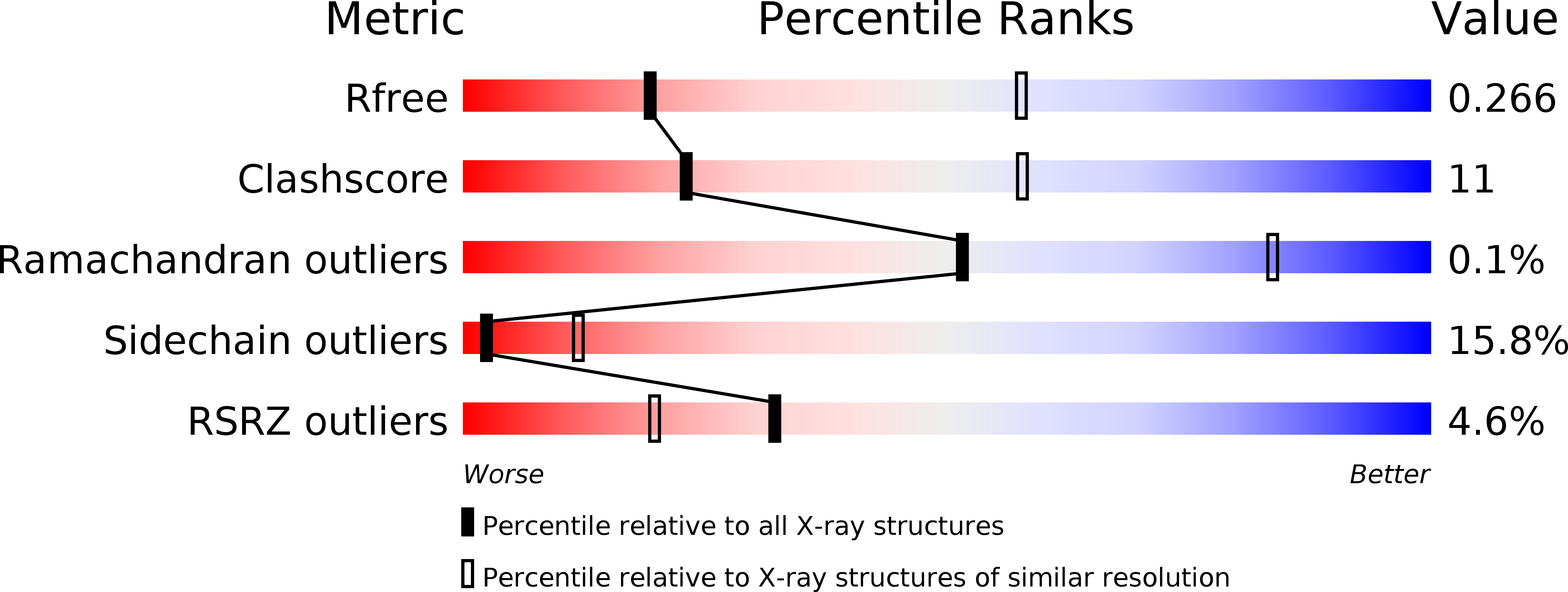
    4KXF

  • PubMed Abstract: 

    Nucleotide-binding and oligomerization domain-like receptor (NLR) proteins oligomerize into multiprotein complexes termed inflammasomes when activated. Their autoinhibition mechanism remains poorly defined. Here, we report the crystal structure of mouse NLRC4 in a closed form. The adenosine diphosphate-mediated interaction between the central nucleotide-binding domain (NBD) and the winged-helix domain (WHD) was critical for stabilizing the closed conformation of NLRC4. The helical domain HD2 repressively contacted a conserved and functionally important α-helix of the NBD. The C-terminal leucine-rich repeat (LRR) domain is positioned to sterically occlude one side of the NBD domain and consequently sequester NLRC4 in a monomeric state. Disruption of ADP-mediated NBD-WHD or NBD-HD2/NBD-LRR interactions resulted in constitutive activation of NLRC4. Together, our data reveal the NBD-organized cooperative autoinhibition mechanism of NLRC4 and provide insight into its activation.


  • Organizational Affiliation

    School of Life Sciences, Tsinghua University, and Tsinghua-Peking Center for Life Sciences, Beijing 100084, China.


Macromolecules
Find similar proteins by:  (by identity cutoff)  |  3D Structure
Entity ID: 1
MoleculeChains Sequence LengthOrganismDetailsImage
NLR family CARD domain-containing protein 41,024Mus musculusMutation(s): 0 
Gene Names: Nlrc4Card12Ipaf
UniProt
Find proteins for Q3UP24 (Mus musculus)
Explore Q3UP24 
Go to UniProtKB:  Q3UP24
Entity Groups  
Sequence Clusters30% Identity50% Identity70% Identity90% Identity95% Identity100% Identity
UniProt GroupQ3UP24
Sequence Annotations
Expand
  • Reference Sequence
Small Molecules
Ligands 2 Unique
IDChains Name / Formula / InChI Key2D Diagram3D Interactions
ADP
Query on ADP

Download Ideal Coordinates CCD File 
I [auth K]
J [auth B]
L [auth D]
M [auth F]
N [auth H]
I [auth K],
J [auth B],
L [auth D],
M [auth F],
N [auth H],
O [auth L],
P [auth N],
R [auth P]
ADENOSINE-5'-DIPHOSPHATE
C10 H15 N5 O10 P2
XTWYTFMLZFPYCI-KQYNXXCUSA-N
SO4
Query on SO4

Download Ideal Coordinates CCD File 
K [auth B],
Q [auth N]
SULFATE ION
O4 S
QAOWNCQODCNURD-UHFFFAOYSA-L
Modified Residues  1 Unique
IDChains TypeFormula2D DiagramParent
SEP
Query on SEP
A [auth K]
B
C [auth D]
D [auth F]
E [auth H]
A [auth K],
B,
C [auth D],
D [auth F],
E [auth H],
F [auth L],
G [auth N],
H [auth P]
L-PEPTIDE LINKINGC3 H8 N O6 PSER
Experimental Data & Validation

Experimental Data

  • Method: X-RAY DIFFRACTION
  • Resolution: 3.20 Å
  • R-Value Free: 0.266 
  • R-Value Work: 0.226 
  • R-Value Observed: 0.228 
  • Space Group: P 41 21 2
Unit Cell:
Length ( Å )Angle ( ˚ )
a = 334.052α = 90
b = 334.052β = 90
c = 177.567γ = 90
Software Package:
Software NamePurpose
HKL-2000data collection
SOLVEphasing
PHENIXrefinement
HKL-2000data reduction
HKL-2000data scaling

Structure Validation

View Full Validation Report


Ligand Structure Quality Assessment 


Entry History 

Deposition Data

Revision History  (Full details and data files)

  • Version 1.0: 2013-07-24
    Type: Initial release