4KP0

Crystal Structure of the human Chymase with TJK002


Experimental Data Snapshot

  • Method: X-RAY DIFFRACTION
  • Resolution: 2.80 Å
  • R-Value Free: 0.330 
  • R-Value Work: 0.226 
  • R-Value Observed: 0.241 

wwPDB Validation   3D Report Full Report


Ligand Structure Quality Assessment 


This is version 1.1 of the entry. See complete history


Literature

Crystal structure of a complex of human chymase with its benzimidazole derived inhibitor

Matsumoto, Y.Kakuda, S.Koizumi, M.Mizuno, T.Muroga, Y.Kawamura, T.Takimoto-Kamimura, M.

(2013) J Synchrotron Radiat 20: 914-918

  • DOI: https://doi.org/10.1107/S0909049513020748
  • Primary Citation of Related Structures:  
    4KP0

  • PubMed Abstract: 

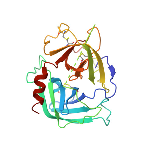
    The crystal structure of human chymase complexed with a novel benzimidazole inhibitor, TJK002, was determined at 2.8 Å resolution. The X-ray crystallographic study shows that the benzimidazole inhibitor forms a non-covalent interaction with the catalytic domain of human chymase. The hydrophobic fragment of the inhibitor occupies the S1 pocket. The carboxylic acid group of the inhibitor forms hydrogen bonds with the imidazole N(ℇ) atom of His57 and/or the O(γ) atom of Ser195 which are members of the catalytic triad. This imidazole ring of His57 induces π-π stacking to the benzene ring of the benzimidazole scaffold as P2 moiety. Fragment molecular orbital calculation of the atomic coordinates by X-ray crystallography shows that this imidazole ring of His57 could be protonated with the carboxyl group of Asp102 or hydroxyl group of Ser195 and the stacking interaction is stabilized. A new drug design strategy is proposed where the stacking to the protonated imidazole of the drug target protein with the benzimidazole scaffold inhibitor causes unpredicted potent inhibitory activity for some enzymes.


  • Organizational Affiliation

    Teijin Institute for Bio-medical Research, 4-3-2 Asahigaoka, Hino, Tokyo 191-8512, Japan.


Macromolecules
Find similar proteins by:  (by identity cutoff)  |  3D Structure
Entity ID: 1
MoleculeChains Sequence LengthOrganismDetailsImage
Chymase226Homo sapiensMutation(s): 0 
Gene Names: CMA1
EC: 3.4.21.39
UniProt & NIH Common Fund Data Resources
Find proteins for P23946 (Homo sapiens)
Explore P23946 
Go to UniProtKB:  P23946
PHAROS:  P23946
GTEx:  ENSG00000092009 
Entity Groups  
Sequence Clusters30% Identity50% Identity70% Identity90% Identity95% Identity100% Identity
UniProt GroupP23946
Sequence Annotations
Expand
  • Reference Sequence
Small Molecules
Ligands 1 Unique
IDChains Name / Formula / InChI Key2D Diagram3D Interactions
KPK
Query on KPK

Download Ideal Coordinates CCD File 
B [auth A]4-({1-[(4-methyl-1-benzothiophen-3-yl)methyl]-1H-benzimidazol-2-yl}sulfanyl)butanoic acid
C21 H20 N2 O2 S2
YSOMBHFITHOMPW-UHFFFAOYSA-N
Binding Affinity Annotations 
IDSourceBinding Affinity
KPK PDBBind:  4KP0 Ki: 2.24 (nM) from 1 assay(s)
BindingDB:  4KP0 IC50: 22 (nM) from 1 assay(s)
Experimental Data & Validation

Experimental Data

  • Method: X-RAY DIFFRACTION
  • Resolution: 2.80 Å
  • R-Value Free: 0.330 
  • R-Value Work: 0.226 
  • R-Value Observed: 0.241 
  • Space Group: P 43 21 2
Unit Cell:
Length ( Å )Angle ( ˚ )
a = 56.92α = 90
b = 56.92β = 90
c = 177.49γ = 90
Software Package:
Software NamePurpose
HKL-2000data collection
MOLREPphasing
REFMACrefinement
HKL-2000data reduction
HKL-2000data scaling

Structure Validation

View Full Validation Report


Ligand Structure Quality Assessment 


Entry History 

Deposition Data

Revision History  (Full details and data files)

  • Version 1.0: 2013-10-30
    Type: Initial release
  • Version 1.1: 2023-11-08
    Changes: Data collection, Database references, Derived calculations, Refinement description