4K71

Crystal structure of a high affinity Human Serum Albumin variant bound to the Neonatal Fc Receptor


Experimental Data Snapshot

  • Method: X-RAY DIFFRACTION
  • Resolution: 2.40 Å
  • R-Value Free: 0.255 
  • R-Value Work: 0.212 
  • R-Value Observed: 0.214 

wwPDB Validation   3D Report Full Report


This is version 1.1 of the entry. See complete history


Literature

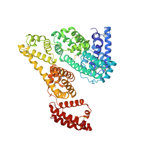
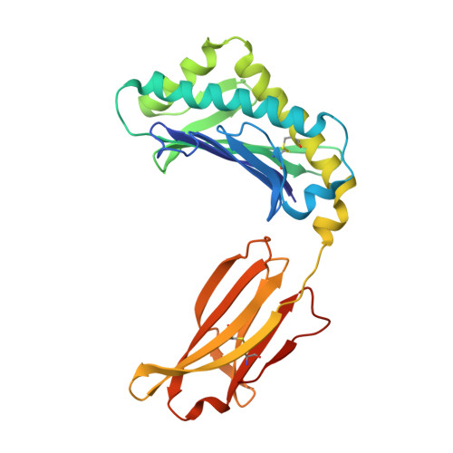
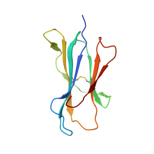
Crystal structure of an HSA/FcRn complex reveals recycling by competitive mimicry of HSA ligands at a pH-dependent hydrophobic interface.

Schmidt, M.M.Townson, S.A.Andreucci, A.J.King, B.M.Schirmer, E.B.Murillo, A.J.Dombrowski, C.Tisdale, A.W.Lowden, P.A.Masci, A.L.Kovalchin, J.T.Erbe, D.V.Wittrup, K.D.Furfine, E.S.Barnes, T.M.

(2013) Structure 21: 1966-1978

  • DOI: https://doi.org/10.1016/j.str.2013.08.022
  • Primary Citation of Related Structures:  
    4K71

  • PubMed Abstract: 

    The long circulating half-life of serum albumin, the most abundant protein in mammalian plasma, derives from pH-dependent endosomal salvage from degradation, mediated by the neonatal Fc receptor (FcRn). Using yeast display, we identified human serum albumin (HSA) variants with increased affinity for human FcRn at endosomal pH, enabling us to solve the crystal structure of a variant HSA/FcRn complex. We find an extensive, primarily hydrophobic interface stabilized by hydrogen-bonding networks involving protonated histidines internal to each protein. The interface features two key FcRn tryptophan side chains inserting into deep hydrophobic pockets on HSA that overlap albumin ligand binding sites. We find that fatty acids (FAs) compete with FcRn, revealing a clash between ligand binding and recycling, and that our high-affinity HSA variants have significantly increased circulating half-lives in mice and monkeys. These observations open the way for the creation of biotherapeutics with significantly improved pharmacokinetics.


  • Organizational Affiliation

    Eleven Biotherapeutics, Inc., Cambridge, MA 02142, USA.


Macromolecules
Find similar proteins by:  (by identity cutoff)  |  3D Structure
Entity ID: 1
MoleculeChains Sequence LengthOrganismDetailsImage
Serum albumin
A, D
585Homo sapiensMutation(s): 4 
Gene Names: ALBGIG20GIG42PRO0903PRO1708PRO2044PRO2619PRO2675UNQ696/PRO1341
UniProt & NIH Common Fund Data Resources
Find proteins for P02768 (Homo sapiens)
Explore P02768 
Go to UniProtKB:  P02768
PHAROS:  P02768
GTEx:  ENSG00000163631 
Entity Groups  
Sequence Clusters30% Identity50% Identity70% Identity90% Identity95% Identity100% Identity
UniProt GroupP02768
Sequence Annotations
Expand
  • Reference Sequence
Find similar proteins by:  (by identity cutoff)  |  3D Structure
Entity ID: 2
MoleculeChains Sequence LengthOrganismDetailsImage
IgG receptor FcRn large subunit p51
B, E
274Homo sapiensMutation(s): 0 
Gene Names: FCGRTFCRN
UniProt & NIH Common Fund Data Resources
Find proteins for P55899 (Homo sapiens)
Explore P55899 
Go to UniProtKB:  P55899
PHAROS:  P55899
GTEx:  ENSG00000104870 
Entity Groups  
Sequence Clusters30% Identity50% Identity70% Identity90% Identity95% Identity100% Identity
UniProt GroupP55899
Sequence Annotations
Expand
  • Reference Sequence
Find similar proteins by:  (by identity cutoff)  |  3D Structure
Entity ID: 3
MoleculeChains Sequence LengthOrganismDetailsImage
Beta-2-microglobulin
C, F
99Homo sapiensMutation(s): 0 
Gene Names: B2MCDABP0092HDCMA22P
UniProt & NIH Common Fund Data Resources
Find proteins for P61769 (Homo sapiens)
Explore P61769 
Go to UniProtKB:  P61769
PHAROS:  P61769
GTEx:  ENSG00000166710 
Entity Groups  
Sequence Clusters30% Identity50% Identity70% Identity90% Identity95% Identity100% Identity
UniProt GroupP61769
Sequence Annotations
Expand
  • Reference Sequence
Small Molecules
Ligands 1 Unique
IDChains Name / Formula / InChI Key2D Diagram3D Interactions
SO4
Query on SO4

Download Ideal Coordinates CCD File 
G [auth A]
H [auth A]
I [auth A]
J [auth A]
K [auth A]
G [auth A],
H [auth A],
I [auth A],
J [auth A],
K [auth A],
L [auth A],
M [auth A],
N [auth B],
O [auth B],
P [auth C],
Q [auth C],
R [auth D],
S [auth D],
T [auth D],
U [auth D],
V [auth D],
W [auth D],
X [auth E],
Y [auth F]
SULFATE ION
O4 S
QAOWNCQODCNURD-UHFFFAOYSA-L
Experimental Data & Validation

Experimental Data

  • Method: X-RAY DIFFRACTION
  • Resolution: 2.40 Å
  • R-Value Free: 0.255 
  • R-Value Work: 0.212 
  • R-Value Observed: 0.214 
  • Space Group: P 21 21 2
Unit Cell:
Length ( Å )Angle ( ˚ )
a = 127.893α = 90
b = 203.536β = 90
c = 100.561γ = 90
Software Package:
Software NamePurpose
HKL-2000data collection
PHASERphasing
REFMACrefinement
XDSdata reduction
XSCALEdata scaling

Structure Validation

View Full Validation Report


Entry History 

Revision History  (Full details and data files)

  • Version 1.0: 2013-10-23
    Type: Initial release
  • Version 1.1: 2014-02-05
    Changes: Database references