4K3X

Crystal structure of a subtype H18 hemagglutinin homologue from A/flat-faced bat/Peru/033/2010 (H18N11)


Experimental Data Snapshot

  • Method: X-RAY DIFFRACTION
  • Resolution: 2.15 Å
  • R-Value Free: 0.220 
  • R-Value Work: 0.183 
  • R-Value Observed: 0.185 

wwPDB Validation   3D Report Full Report


Ligand Structure Quality Assessment 


This is version 2.1 of the entry. See complete history


Literature

New world bats harbor diverse influenza a viruses.

Tong, S.Zhu, X.Li, Y.Shi, M.Zhang, J.Bourgeois, M.Yang, H.Chen, X.Recuenco, S.Gomez, J.Chen, L.M.Johnson, A.Tao, Y.Dreyfus, C.Yu, W.McBride, R.Carney, P.J.Gilbert, A.T.Chang, J.Guo, Z.Davis, C.T.Paulson, J.C.Stevens, J.Rupprecht, C.E.Holmes, E.C.Wilson, I.A.Donis, R.O.

(2013) PLoS Pathog 9: e1003657-e1003657

  • DOI: https://doi.org/10.1371/journal.ppat.1003657
  • Primary Citation of Related Structures:  
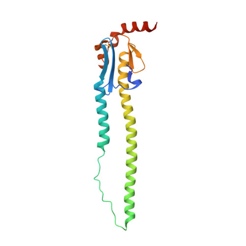
    4K3X, 4K3Y, 4MC5, 4MC7

  • PubMed Abstract: 

    Aquatic birds harbor diverse influenza A viruses and are a major viral reservoir in nature. The recent discovery of influenza viruses of a new H17N10 subtype in Central American fruit bats suggests that other New World species may similarly carry divergent influenza viruses. Using consensus degenerate RT-PCR, we identified a novel influenza A virus, designated as H18N11, in a flat-faced fruit bat (Artibeus planirostris) from Peru. Serologic studies with the recombinant H18 protein indicated that several Peruvian bat species were infected by this virus. Phylogenetic analyses demonstrate that, in some gene segments, New World bats harbor more influenza virus genetic diversity than all other mammalian and avian species combined, indicative of a long-standing host-virus association. Structural and functional analyses of the hemagglutinin and neuraminidase indicate that sialic acid is not a ligand for virus attachment nor a substrate for release, suggesting a unique mode of influenza A virus attachment and activation of membrane fusion for entry into host cells. Taken together, these findings indicate that bats constitute a potentially important and likely ancient reservoir for a diverse pool of influenza viruses.


  • Organizational Affiliation

    Division of Viral Diseases, Centers for Disease Control and Prevention, Atlanta, Georgia, United States of America.


Macromolecules
Find similar proteins by:  (by identity cutoff)  |  3D Structure
Entity ID: 1
MoleculeChains Sequence LengthOrganismDetailsImage
Hemagglutinin HA1A,
B [auth C],
C [auth E]
329Influenza A virusMutation(s): 0 
Entity Groups  
Sequence Clusters30% Identity50% Identity70% Identity90% Identity95% Identity100% Identity
Sequence Annotations
Expand
  • Reference Sequence
Find similar proteins by:  (by identity cutoff)  |  3D Structure
Entity ID: 2
MoleculeChains Sequence LengthOrganismDetailsImage
Hemagglutinin HA2D [auth B],
E [auth D],
F
181Influenza A virusMutation(s): 0 
Entity Groups  
Sequence Clusters30% Identity50% Identity70% Identity90% Identity95% Identity100% Identity
Sequence Annotations
Expand
  • Reference Sequence
Oligosaccharides

Help

Entity ID: 3
MoleculeChains Length2D Diagram Glycosylation3D Interactions
alpha-D-mannopyranose-(1-3)-[alpha-D-mannopyranose-(1-6)]beta-D-mannopyranose-(1-4)-2-acetamido-2-deoxy-beta-D-glucopyranose-(1-4)-[beta-L-fucopyranose-(1-3)][alpha-L-fucopyranose-(1-6)]2-acetamido-2-deoxy-beta-D-glucopyranose
G, H, K
7N-Glycosylation
Glycosylation Resources
GlyTouCan:  G87407JY
GlyCosmos:  G87407JY
GlyGen:  G87407JY
Entity ID: 4
MoleculeChains Length2D Diagram Glycosylation3D Interactions
beta-L-fucopyranose-(1-3)-[2-acetamido-2-deoxy-beta-D-glucopyranose-(1-4)][beta-L-fucopyranose-(1-6)]2-acetamido-2-deoxy-beta-D-glucopyranose
I
4N-Glycosylation
Glycosylation Resources
GlyTouCan:  G44123WW
GlyCosmos:  G44123WW
Entity ID: 5
MoleculeChains Length2D Diagram Glycosylation3D Interactions
2-acetamido-2-deoxy-beta-D-glucopyranose-(1-4)-2-acetamido-2-deoxy-beta-D-glucopyranose
J, M, O, Q, R
2N-Glycosylation
Glycosylation Resources
GlyTouCan:  G42666HT
GlyCosmos:  G42666HT
GlyGen:  G42666HT
Entity ID: 6
MoleculeChains Length2D Diagram Glycosylation3D Interactions
beta-D-mannopyranose-(1-6)-beta-D-mannopyranose-(1-4)-2-acetamido-2-deoxy-beta-D-glucopyranose-(1-4)-[beta-L-fucopyranose-(1-6)]2-acetamido-2-deoxy-beta-D-glucopyranose
L
5N-Glycosylation
Glycosylation Resources
GlyTouCan:  G81574IL
GlyCosmos:  G81574IL
GlyGen:  G81574IL
Entity ID: 7
MoleculeChains Length2D Diagram Glycosylation3D Interactions
beta-L-fucopyranose-(1-3)-[2-acetamido-2-deoxy-beta-D-glucopyranose-(1-4)][alpha-L-fucopyranose-(1-6)]2-acetamido-2-deoxy-beta-D-glucopyranose
N
4N-Glycosylation
Glycosylation Resources
GlyTouCan:  G62810GW
GlyCosmos:  G62810GW
Entity ID: 8
MoleculeChains Length2D Diagram Glycosylation3D Interactions
beta-L-fucopyranose-(1-3)-[2-acetamido-2-deoxy-beta-D-glucopyranose-(1-4)]2-acetamido-2-deoxy-beta-D-glucopyranose
P
3N-Glycosylation
Glycosylation Resources
GlyTouCan:  G46874CY
GlyCosmos:  G46874CY
GlyGen:  G46874CY
Small Molecules
Ligands 2 Unique
IDChains Name / Formula / InChI Key2D Diagram3D Interactions
NAG
Query on NAG

Download Ideal Coordinates CCD File 
T [auth C],
X [auth E]
2-acetamido-2-deoxy-beta-D-glucopyranose
C8 H15 N O6
OVRNDRQMDRJTHS-FMDGEEDCSA-N
P4G
Query on P4G

Download Ideal Coordinates CCD File 
AA [auth B]
BA [auth B]
CA [auth B]
DA [auth D]
EA [auth F]
AA [auth B],
BA [auth B],
CA [auth B],
DA [auth D],
EA [auth F],
FA [auth F],
S [auth A],
U [auth C],
V [auth C],
W [auth C],
Y [auth E],
Z [auth E]
1-ETHOXY-2-(2-ETHOXYETHOXY)ETHANE
C8 H18 O3
RRQYJINTUHWNHW-UHFFFAOYSA-N
Experimental Data & Validation

Experimental Data

  • Method: X-RAY DIFFRACTION
  • Resolution: 2.15 Å
  • R-Value Free: 0.220 
  • R-Value Work: 0.183 
  • R-Value Observed: 0.185 
  • Space Group: I 4 2 2
Unit Cell:
Length ( Å )Angle ( ˚ )
a = 238.884α = 90
b = 238.884β = 90
c = 160.785γ = 90
Software Package:
Software NamePurpose
HKL-2000data collection
PHASERphasing
PHENIXrefinement
HKL-2000data reduction
HKL-2000data scaling

Structure Validation

View Full Validation Report


Ligand Structure Quality Assessment 


Entry History 

Deposition Data

Revision History  (Full details and data files)

  • Version 1.0: 2013-10-23
    Type: Initial release
  • Version 1.1: 2013-11-20
    Changes: Database references
  • Version 2.0: 2020-07-29
    Type: Remediation
    Reason: Carbohydrate remediation
    Changes: Advisory, Atomic model, Data collection, Derived calculations, Structure summary
  • Version 2.1: 2023-09-20
    Changes: Data collection, Database references, Refinement description, Structure summary