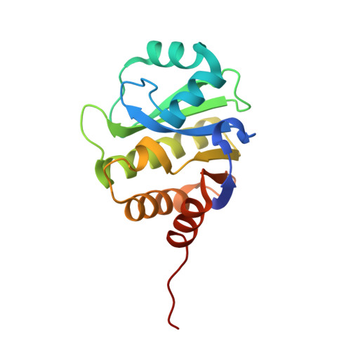
Crystal Structure of the evolved variant of the computationally designed serine hydrolase, Northeast Structural Genomics Consortium (NESG) Target OR275
Kuzin, A., Lew, S., Rajagopalan, S., Seetharaman, J., Mao, L., Xiao, R., Lee, D., Everett, J.K., Acton, T.B., Baker, D., Montelione, G.T., Tong, L., Hunt, J.F.To be published.