4J8R

Structure of an octapeptide repeat of the prion protein bound to the POM2 Fab antibody fragment


Experimental Data Snapshot

  • Method: X-RAY DIFFRACTION
  • Resolution: 2.30 Å
  • R-Value Free: 0.257 
  • R-Value Work: 0.247 
  • R-Value Observed: 0.247 

wwPDB Validation   3D Report Full Report


This is version 1.3 of the entry. See complete history


Literature

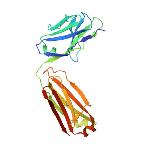
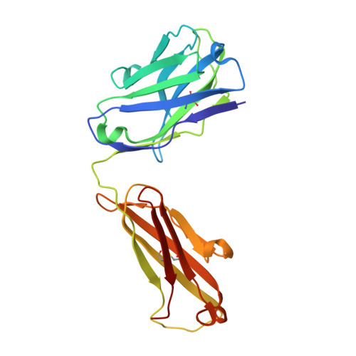
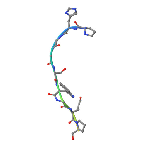
The crystal structure of an octapeptide repeat of the Prion protein in complex with a Fab fragment of the POM2 antibody.

Swayampakula, M.Baral, P.K.Aguzzi, A.Kav, N.N.James, M.N.

(2013) Protein Sci 22: 893-903

  • DOI: https://doi.org/10.1002/pro.2270
  • Primary Citation of Related Structures:  
    4J8R

  • PubMed Abstract: 

    Prion diseases are progressive, infectious neurodegenerative disorders caused primarily by the misfolding of the cellular prion protein (PrP(c)) into an insoluble, protease-resistant, aggregated isoform termed PrP(sc). In native conditions, PrP(c) has a structured C-terminal domain and a highly flexible N-terminal domain. A part of this N-terminal domain consists of 4-5 repeats of an unusual glycine-rich, eight amino acids long peptide known as the octapeptide repeat (OR) domain. In this article, we successfully report the first crystal structure of an OR of PrP(c) bound to the Fab fragment of the POM2 antibody. The structure was solved at a resolution of 2.3 Å by molecular replacement. Although several studies have previously predicted a β-turn-like structure of the unbound ORs, our structure shows an extended conformation of the OR when bound to a molecule of the POM2 Fab indicating that the bound Fab disrupts any putative native β turn conformation of the ORs. Encouraging results from several recent studies have shown that administering small molecule ligands or antibodies targeting the OR domain of PrP result in arresting the progress of peripheral prion infections both in ex vivo and in in vivo models. This makes the structural study of the interactions of POM2 Fab with the OR domain very important as it would help us to design smaller and tighter binding OR ligands.


  • Organizational Affiliation

    Department of Biochemistry, Faculty of Medicine and Dentistry, University of Alberta, Edmonton, Alberta, T6G 2H7, Canada.


Macromolecules
Find similar proteins by:  (by identity cutoff)  |  3D Structure
Entity ID: 1
MoleculeChains Sequence LengthOrganismDetailsImage
Light chain of POM2 FabA,
D [auth C]
210Mus musculusMutation(s): 0 
Entity Groups  
Sequence Clusters30% Identity50% Identity70% Identity90% Identity95% Identity100% Identity
Sequence Annotations
Expand
  • Reference Sequence
Find similar proteins by:  (by identity cutoff)  |  3D Structure
Entity ID: 2
MoleculeChains Sequence LengthOrganismDetailsImage
Heavy chain of POM2 FabB,
E [auth D]
220Mus musculusMutation(s): 0 
Entity Groups  
Sequence Clusters30% Identity50% Identity70% Identity90% Identity95% Identity100% Identity
Sequence Annotations
Expand
  • Reference Sequence

Find similar proteins by:  Sequence   |   3D Structure  

Entity ID: 3
MoleculeChains Sequence LengthOrganismDetailsImage
Major prion proteinC [auth I],
F [auth J]
16Mus musculusMutation(s): 0 
UniProt
Find proteins for P04925 (Mus musculus)
Explore P04925 
Go to UniProtKB:  P04925
Entity Groups  
Sequence Clusters30% Identity50% Identity70% Identity90% Identity95% Identity100% Identity
UniProt GroupP04925
Sequence Annotations
Expand
  • Reference Sequence
Experimental Data & Validation

Experimental Data

  • Method: X-RAY DIFFRACTION
  • Resolution: 2.30 Å
  • R-Value Free: 0.257 
  • R-Value Work: 0.247 
  • R-Value Observed: 0.247 
  • Space Group: P 21 2 21
Unit Cell:
Length ( Å )Angle ( ˚ )
a = 65.415α = 90
b = 71.567β = 90
c = 207.592γ = 90
Software Package:
Software NamePurpose
HKL-2000data collection
PHASERphasing
PHENIXrefinement
autoXDSdata reduction
autoXDSdata scaling

Structure Validation

View Full Validation Report


Entry History 

Deposition Data

Revision History  (Full details and data files)

  • Version 1.0: 2013-05-22
    Type: Initial release
  • Version 1.1: 2013-07-24
    Changes: Database references
  • Version 1.2: 2014-11-26
    Changes: Other
  • Version 1.3: 2023-09-20
    Changes: Data collection, Database references, Refinement description