4J03

Crystal structure of human soluble epoxide hydrolase complexed with fulvestrant


Experimental Data Snapshot

  • Method: X-RAY DIFFRACTION
  • Resolution: 2.92 Å
  • R-Value Free: 0.250 
  • R-Value Work: 0.188 
  • R-Value Observed: 0.191 

wwPDB Validation   3D Report Full Report


Ligand Structure Quality Assessment 


This is version 1.3 of the entry. See complete history


Literature

Inhibition of soluble epoxide hydrolase by fulvestrant and sulfoxides.

Morisseau, C.Pakhomova, S.Hwang, S.H.Newcomer, M.E.Hammock, B.D.

(2013) Bioorg Med Chem Lett 23: 3818-3821

  • DOI: https://doi.org/10.1016/j.bmcl.2013.04.083
  • Primary Citation of Related Structures:  
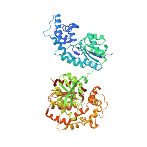
    4J03

  • PubMed Abstract: 

    The soluble epoxide hydrolase (sEH) is a key enzyme in the metabolism of epoxy-fatty acids, signaling molecules involved in numerous biologies. Toward finding novel inhibitors of sEH, a library of known drugs was tested for inhibition of sEH. We found that fulvestrant, an anticancer agent, is a potent (KI=26 nM) competitive inhibitor of sEH. From this observation, we found that alkyl-sulfoxides represent a new kind of pharmacophore for the inhibition of sEH.


  • Organizational Affiliation

    Department of Entomology and UCD Comprehensive Cancer Center, University of California, One Shields Avenue, Davis, CA 95616, USA. chmorisseau@ucdavis.edu


Macromolecules
Find similar proteins by:  (by identity cutoff)  |  3D Structure
Entity ID: 1
MoleculeChains Sequence LengthOrganismDetailsImage
Bifunctional epoxide hydrolase 2555Homo sapiensMutation(s): 0 
Gene Names: EPHX2
EC: 3.3.2.10 (PDB Primary Data), 3.1.3.76 (PDB Primary Data)
UniProt & NIH Common Fund Data Resources
Find proteins for P34913 (Homo sapiens)
Explore P34913 
Go to UniProtKB:  P34913
PHAROS:  P34913
GTEx:  ENSG00000120915 
Entity Groups  
Sequence Clusters30% Identity50% Identity70% Identity90% Identity95% Identity100% Identity
UniProt GroupP34913
Sequence Annotations
Expand
  • Reference Sequence
Small Molecules
Ligands 3 Unique
IDChains Name / Formula / InChI Key2D Diagram3D Interactions
FVS
Query on FVS

Download Ideal Coordinates CCD File 
D [auth A](7beta,9beta,13alpha,17beta)-7-{9-[(R)-(4,4,5,5,5-pentafluoropentyl)sulfinyl]nonyl}estra-1(10),2,4-triene-3,17-diol
C32 H47 F5 O3 S
VWUXBMIQPBEWFH-CIAKRVSBSA-N
PO4
Query on PO4

Download Ideal Coordinates CCD File 
B [auth A]PHOSPHATE ION
O4 P
NBIIXXVUZAFLBC-UHFFFAOYSA-K
MG
Query on MG

Download Ideal Coordinates CCD File 
C [auth A]MAGNESIUM ION
Mg
JLVVSXFLKOJNIY-UHFFFAOYSA-N
Binding Affinity Annotations 
IDSourceBinding Affinity
FVS PDBBind:  4J03 Ki: 26 (nM) from 1 assay(s)
Experimental Data & Validation

Experimental Data

  • Method: X-RAY DIFFRACTION
  • Resolution: 2.92 Å
  • R-Value Free: 0.250 
  • R-Value Work: 0.188 
  • R-Value Observed: 0.191 
  • Space Group: P 65 2 2
Unit Cell:
Length ( Å )Angle ( ˚ )
a = 92.507α = 90
b = 92.507β = 90
c = 243.811γ = 120
Software Package:
Software NamePurpose
MOLREPphasing
REFMACrefinement
HKL-2000data reduction
HKL-2000data scaling

Structure Validation

View Full Validation Report


Ligand Structure Quality Assessment 


Entry History 

Deposition Data

Revision History  (Full details and data files)

  • Version 1.0: 2013-06-05
    Type: Initial release
  • Version 1.1: 2013-06-19
    Changes: Database references
  • Version 1.2: 2013-06-26
    Changes: Database references
  • Version 1.3: 2023-09-20
    Changes: Data collection, Database references, Derived calculations, Refinement description