4IHO

Crystal structure of H-2Db Y159F in complex with chimeric gp100


Experimental Data Snapshot

  • Method: X-RAY DIFFRACTION
  • Resolution: 2.80 Å
  • R-Value Free: 0.305 
  • R-Value Work: 0.249 
  • R-Value Observed: 0.252 

wwPDB Validation   3D Report Full Report


This is version 1.2 of the entry. See complete history


Literature

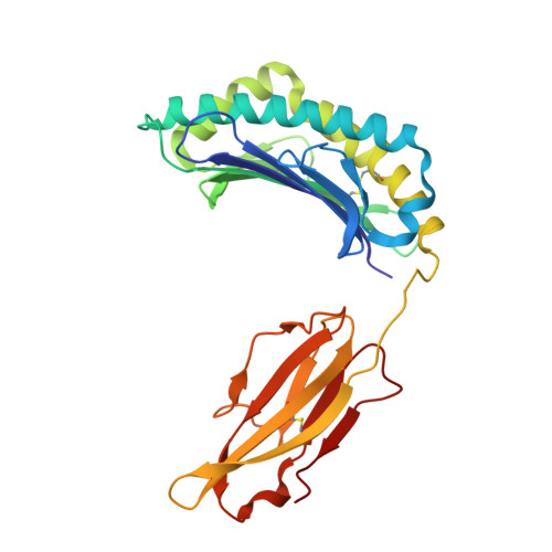
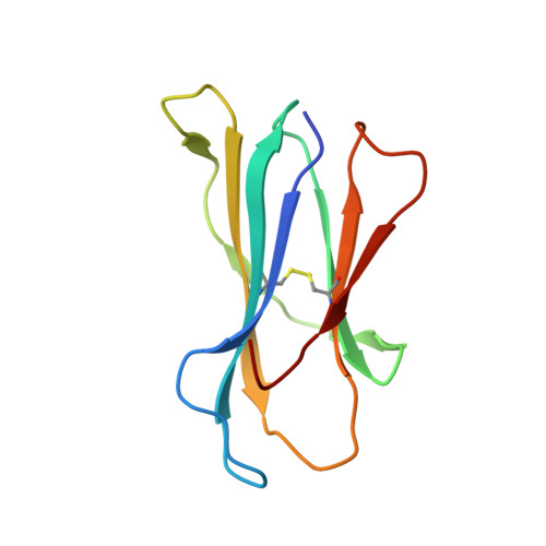
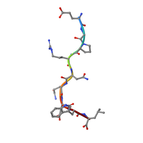
Proline substitution independently enhances H-2D(b) complex stabilization and TCR recognition of melanoma-associated peptides

Uchtenhagen, H.Abualrous, E.T.Stahl, E.Allerbring, E.B.Sluijter, M.Zacharias, M.Sandalova, T.van Hall, T.Springer, S.Nygren, P.A.Achour, A.

(2013) Eur J Immunol 43: 3051-3060

  • DOI: https://doi.org/10.1002/eji.201343456
  • Primary Citation of Related Structures:  
    4IHO

  • PubMed Abstract: 

    The immunogenicity of H-2D(b) (D(b)) restricted epitopes can be significantly increased by substituting peptide position 3 to a proline (p3P). The p3P modification enhances MHC stability without altering the conformation of the modified epitope allowing for T-cell cross-reactivity with the native peptide. The present study reveals how specific interactions between p3P and the highly conserved MHC heavy chain residue Y159 increase the stability of D(b) in complex with an optimized version of the melanoma-associated epitope gp10025-33 . Furthermore, the p3P modification directly increased the affinity of the D(b)/gp10025-33 -specific T-cell receptor (TCR) pMel. Surprisingly, the enhanced TCR binding was independent from the observed increased stability of the optimized D(b)/gp10025-33 complex and from the interactions formed between p3P and Y159, indicating a direct effect of the p3P modification on TCR recognition.


  • Organizational Affiliation

    Science for Life Laboratory, Center for Infectious Medicine (CIM), Department of Medicine, Karolinska Insitutet, Karolinska University Hospital Huddinge, Stockholm, Sweden.


Macromolecules
Find similar proteins by:  (by identity cutoff)  |  3D Structure
Entity ID: 1
MoleculeChains Sequence LengthOrganismDetailsImage
H-2 class I histocompatibility antigen, D-B alpha chain
A, D
276Mus musculusMutation(s): 1 
Gene Names: H2-D1
UniProt
Find proteins for P01899 (Mus musculus)
Explore P01899 
Go to UniProtKB:  P01899
Entity Groups  
Sequence Clusters30% Identity50% Identity70% Identity90% Identity95% Identity100% Identity
UniProt GroupP01899
Sequence Annotations
Expand
  • Reference Sequence
Find similar proteins by:  (by identity cutoff)  |  3D Structure
Entity ID: 2
MoleculeChains Sequence LengthOrganismDetailsImage
Beta-2-microglobulin
B, E
99Mus musculusMutation(s): 0 
Gene Names: B2m
UniProt & NIH Common Fund Data Resources
Find proteins for P01887 (Mus musculus)
Explore P01887 
Go to UniProtKB:  P01887
IMPC:  MGI:88127
Entity Groups  
Sequence Clusters30% Identity50% Identity70% Identity90% Identity95% Identity100% Identity
UniProt GroupP01887
Sequence Annotations
Expand
  • Reference Sequence

Find similar proteins by:  Sequence   |   3D Structure  

Entity ID: 3
MoleculeChains Sequence LengthOrganismDetailsImage
NONAMERIC PEPTIDE CHIMERIC GP100
C, F
9N/AMutation(s): 0 
Sequence Annotations
Expand
  • Reference Sequence
Experimental Data & Validation

Experimental Data

  • Method: X-RAY DIFFRACTION
  • Resolution: 2.80 Å
  • R-Value Free: 0.305 
  • R-Value Work: 0.249 
  • R-Value Observed: 0.252 
  • Space Group: I 2 2 2
Unit Cell:
Length ( Å )Angle ( ˚ )
a = 90.661α = 90
b = 146.011β = 90
c = 187.585γ = 90
Software Package:
Software NamePurpose
ADSCdata collection
PHASERphasing
REFMACrefinement
XDSdata reduction
SCALAdata scaling

Structure Validation

View Full Validation Report


Entry History 

Deposition Data

Revision History  (Full details and data files)

  • Version 1.0: 2013-09-11
    Type: Initial release
  • Version 1.1: 2013-12-11
    Changes: Database references
  • Version 1.2: 2023-11-08
    Changes: Data collection, Database references, Derived calculations, Refinement description