4I9W

Human two pore domain K+ channel TRAAK (K2P4.1) - Fab complex structure


Experimental Data Snapshot

  • Method: X-RAY DIFFRACTION
  • Resolution: 2.75 Å
  • R-Value Free: 0.236 
  • R-Value Work: 0.201 
  • R-Value Observed: 0.203 

wwPDB Validation   3D Report Full Report


This is version 1.2 of the entry. See complete history


Literature

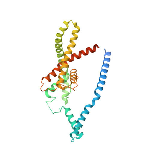
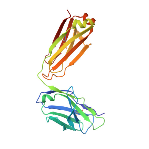
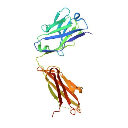
Domain-swapped chain connectivity and gated membrane access in a Fab-mediated crystal of the human TRAAK K+ channel.

Brohawn, S.G.Campbell, E.B.Mackinnon, R.

(2013) Proc Natl Acad Sci U S A 110: 2129-2134

  • DOI: https://doi.org/10.1073/pnas.1218950110
  • Primary Citation of Related Structures:  
    4I9W

  • PubMed Abstract: 

    TRAAK (TWIK-related arachidonic acid-stimulated K(+) channel, K2P4.1) K(+) ion channels are expressed predominantly in the nervous system to control cellular resting membrane potential and are regulated by mechanical and chemical properties of the lipid membrane. TRAAK channels are twofold symmetric, which precludes a direct extension of gating mechanisms that close canonical fourfold symmetric K(+) channels. We present the crystal structure of human TRAAK in complex with antibody antigen-binding fragments (Fabs) at 2.75-Å resolution. In contrast to a previous structure, this structure reveals a domain-swapped chain connectivity enabled by the helical cap that exchanges two opposing outer helices 180° around the channel. An unrelated conformational change of an inner helix seals a side opening to the membrane bilayer and is associated with structural changes around the K(+)-selectivity filter that may have implications for mechanosensitivity and gating of TRAAK channels.


  • Organizational Affiliation

    Laboratory of Molecular Neurobiology and Biophysics and Howard Hughes Medical Institute, The Rockefeller University, New York, NY 10065, USA.


Macromolecules
Find similar proteins by:  (by identity cutoff)  |  3D Structure
Entity ID: 1
MoleculeChains Sequence LengthOrganismDetailsImage
Potassium channel subfamily K member 4
A, B
309Homo sapiensMutation(s): 2 
Gene Names: K2P4.1KCNK4TRAAK
Membrane Entity: Yes 
UniProt & NIH Common Fund Data Resources
Find proteins for Q9NYG8 (Homo sapiens)
Explore Q9NYG8 
Go to UniProtKB:  Q9NYG8
PHAROS:  Q9NYG8
GTEx:  ENSG00000182450 
Entity Groups  
Sequence Clusters30% Identity50% Identity70% Identity90% Identity95% Identity100% Identity
UniProt GroupQ9NYG8
Sequence Annotations
Expand
  • Reference Sequence
Find similar proteins by:  (by identity cutoff)  |  3D Structure
Entity ID: 2
MoleculeChains Sequence LengthOrganismDetailsImage
ANTIBODY FAB FRAGMENT LIGHT CHAINC [auth D],
E [auth F]
211Mus musculusMutation(s): 0 
Entity Groups  
Sequence Clusters30% Identity50% Identity70% Identity90% Identity95% Identity100% Identity
Sequence Annotations
Expand
  • Reference Sequence
Find similar proteins by:  (by identity cutoff)  |  3D Structure
Entity ID: 3
MoleculeChains Sequence LengthOrganismDetailsImage
ANTIBODY FAB FRAGMENT HEAVY CHAIND [auth E],
F [auth G]
217Mus musculusMutation(s): 0 
Entity Groups  
Sequence Clusters30% Identity50% Identity70% Identity90% Identity95% Identity100% Identity
Sequence Annotations
Expand
  • Reference Sequence
Experimental Data & Validation

Experimental Data

  • Method: X-RAY DIFFRACTION
  • Resolution: 2.75 Å
  • R-Value Free: 0.236 
  • R-Value Work: 0.201 
  • R-Value Observed: 0.203 
  • Space Group: P 1 21 1
Unit Cell:
Length ( Å )Angle ( ˚ )
a = 80.887α = 90
b = 138.905β = 94.87
c = 97.461γ = 90
Software Package:
Software NamePurpose
HKL-2000data collection
PHASERphasing
REFMACrefinement
HKL-2000data reduction
HKL-2000data scaling

Structure Validation

View Full Validation Report


Entry History 

Deposition Data

Revision History  (Full details and data files)

  • Version 1.0: 2013-01-23
    Type: Initial release
  • Version 1.1: 2013-02-20
    Changes: Database references
  • Version 1.2: 2023-09-20
    Changes: Data collection, Database references, Derived calculations, Refinement description