


Iodide SAD phased crystal structure of a phosphoglucomutase from Brucella melitensis complexed with glucose-6-phosphate
Fairman, J.W., Craig, T.K., Staker, B.L.To be published.
Experimental Data Snapshot
Entity ID: 1 | |||||
|---|---|---|---|---|---|
| Molecule | Chains | Sequence Length | Organism | Details | Image |
| Phosphomannomutase | 481 | Brucella melitensis bv. 1 str. 16M | Mutation(s): 0 Gene Names: BAWG_1686 EC: 5.4.2.8 | 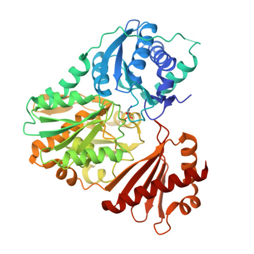 | |
Entity Groups | |||||
| Sequence Clusters | 30% Identity50% Identity70% Identity90% Identity95% Identity100% Identity | ||||
Sequence AnnotationsExpand | |||||
| |||||
| Ligands 4 Unique | |||||
|---|---|---|---|---|---|
| ID | Chains | Name / Formula / InChI Key | 2D Diagram | 3D Interactions | |
| G6Q Query on G6Q | IC [auth B], TA [auth A] | GLUCOSE-6-PHOSPHATE C6 H13 O9 P VFRROHXSMXFLSN-SLPGGIOYSA-N |  | ||
| IOD Query on IOD | AA [auth A] AC [auth B] BA [auth A] BC [auth B] C [auth A] | IODIDE ION I XMBWDFGMSWQBCA-UHFFFAOYSA-M |  | ||
| EDO Query on EDO | AB [auth A] JC [auth B] UA [auth A] VA [auth A] WA [auth A] | 1,2-ETHANEDIOL C2 H6 O2 LYCAIKOWRPUZTN-UHFFFAOYSA-N |  | ||
| MG Query on MG | BB [auth A], KC [auth B] | MAGNESIUM ION Mg JLVVSXFLKOJNIY-UHFFFAOYSA-N |  | ||
| Modified Residues 1 Unique | |||||
|---|---|---|---|---|---|
| ID | Chains | Type | Formula | 2D Diagram | Parent |
| SEP Query on SEP | A, B | L-PEPTIDE LINKING | C3 H8 N O6 P |  | SER |
| Length ( Å ) | Angle ( ˚ ) |
|---|---|
| a = 61.95 | α = 90 |
| b = 55.98 | β = 92.18 |
| c = 131.67 | γ = 90 |
| Software Name | Purpose |
|---|---|
| XSCALE | data scaling |
| REFMAC | refinement |
| PDB_EXTRACT | data extraction |
| PHASER | phasing |
RCSB PDB (citation) is hosted by
RCSB PDB is a member of the

