4H4J

Crystal structure of a N-acetylmuramoyl-L-alanine amidase (BACUNI_02947) from Bacteroides uniformis ATCC 8492 at 1.15 A resolution


Experimental Data Snapshot

  • Method: X-RAY DIFFRACTION
  • Resolution: 1.15 Å
  • R-Value Free: 0.164 
  • R-Value Work: 0.131 
  • R-Value Observed: 0.132 

wwPDB Validation   3D Report Full Report


This is version 1.5 of the entry. See complete history


Literature

Structure-guided functional characterization of DUF1460 reveals a highly specific NlpC/P60 amidase family.

Xu, Q.Mengin-Lecreulx, D.Patin, D.Grant, J.C.Chiu, H.J.Jaroszewski, L.Knuth, M.W.Godzik, A.Lesley, S.A.Elsliger, M.A.Deacon, A.M.Wilson, I.A.

(2014) Structure 22: 1799-1809

  • DOI: https://doi.org/10.1016/j.str.2014.09.018
  • Primary Citation of Related Structures:  
    4H4J, 4Q5K, 4Q68

  • PubMed Abstract: 

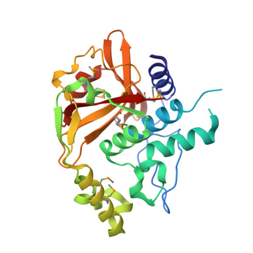
    GlcNAc-1,6-anhydro-MurNAc-tetrapeptide is a major peptidoglycan degradation intermediate and a cytotoxin. It is generated by lytic transglycosylases and further degraded and recycled by various enzymes. We have identified and characterized a highly specific N-acetylmuramoyl-L-alanine amidase (AmiA) from Bacteroides uniformis, a member of the DUF1460 protein family, that hydrolyzes GlcNAc-1,6-anhydro-MurNAc-peptide into disaccharide and stem peptide. The high-resolution apo structure at 1.15 Å resolution shows that AmiA is related to NlpC/P60 γ-D-Glu-meso-diaminopimelic acid amidases and shares a common catalytic core and cysteine peptidase-like active site. AmiA has evolved structural adaptations that reconfigure the substrate recognition site. The preferred substrates for AmiA were predicted in silico based on structural and bioinformatics data, and subsequently were characterized experimentally. Further crystal structures of AmiA in complexes with GlcNAc-1,6-anhydro-MurNAc and GlcNAc have enabled us to elucidate substrate recognition and specificity. DUF1460 is highly conserved in structure and defines another amidase family.


  • Organizational Affiliation

    Joint Center for Structural Genomics (JCSG); Stanford Synchrotron Radiation Lightsource, SLAC National Accelerator Laboratory, Menlo Park, CA 94025, USA.


Macromolecules
Find similar proteins by:  (by identity cutoff)  |  3D Structure
Entity ID: 1
MoleculeChains Sequence LengthOrganismDetailsImage
hypothetical protein240Bacteroides uniformis ATCC 8492Mutation(s): 0 
Gene Names: BACUNI_02947ZP_02071508.1
UniProt
Find proteins for A7V5T8 (Bacteroides uniformis (strain ATCC 8492 / DSM 6597 / CCUG 4942 / CIP 103695 / JCM 5828 / KCTC 5204 / NCTC 13054 / VPI 0061))
Explore A7V5T8 
Go to UniProtKB:  A7V5T8
Entity Groups  
Sequence Clusters30% Identity50% Identity70% Identity90% Identity95% Identity100% Identity
UniProt GroupA7V5T8
Sequence Annotations
Expand
  • Reference Sequence
Small Molecules
Modified Residues  1 Unique
IDChains TypeFormula2D DiagramParent
MSE
Query on MSE
A
L-PEPTIDE LINKINGC5 H11 N O2 SeMET
Experimental Data & Validation

Experimental Data

Unit Cell:
Length ( Å )Angle ( ˚ )
a = 46.349α = 90
b = 63.713β = 90
c = 72.962γ = 90
Software Package:
Software NamePurpose
MolProbitymodel building
PDB_EXTRACTdata extraction
SHELXphasing
SHARPphasing
XSCALEdata scaling
REFMACrefinement
XDSdata reduction
SHELXDphasing

Structure Validation

View Full Validation Report


Entry History 

Deposition Data

Revision History  (Full details and data files)

  • Version 1.0: 2012-10-17
    Type: Initial release
  • Version 1.1: 2014-12-24
    Changes: Structure summary
  • Version 1.2: 2017-11-15
    Changes: Refinement description
  • Version 1.3: 2018-01-24
    Changes: Database references
  • Version 1.4: 2019-09-18
    Changes: Data collection, Database references, Derived calculations
  • Version 1.5: 2023-02-01
    Changes: Database references