4GXQ

Crystal Structure of ATP bound RpMatB-BxBclM chimera B1


Experimental Data Snapshot

  • Method: X-RAY DIFFRACTION
  • Resolution: 2.00 Å
  • R-Value Free: 0.254 
  • R-Value Work: 0.219 
  • R-Value Observed: 0.221 

wwPDB Validation   3D Report Full Report


Ligand Structure Quality Assessment 


This is version 1.3 of the entry. See complete history


Literature

Structural Insights into the Substrate Specificity of the Rhodopseudomonas palustris Protein Acetyltransferase RpPat: IDENTIFICATION OF A LOOP CRITICAL FOR RECOGNITION BY RpPat.

Crosby, H.A.Rank, K.C.Rayment, I.Escalante-Semerena, J.C.

(2012) J Biol Chem 287: 41392-41404

  • DOI: https://doi.org/10.1074/jbc.M112.417360
  • Primary Citation of Related Structures:  
    4GXQ, 4GXR

  • PubMed Abstract: 

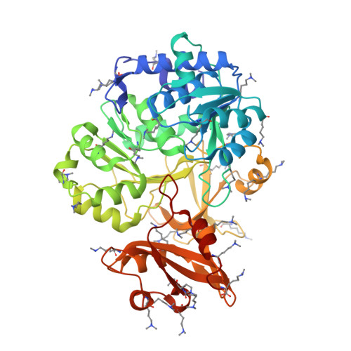
    Lysine acetylation is a post-translational modification that is important for the regulation of metabolism in both prokaryotes and eukaryotes. In bacteria, the best studied protein acetyltransferase is Pat. In the purple photosynthetic bacterium Rhodopseudomonas palustris, at least 10 AMP-forming acyl-CoA synthetase enzymes are acetylated by the Pat homologue RpPat. All bona fide RpPat substrates contain the conserved motif PX(4)GK. Here, we show that the presence of such a motif is necessary but not sufficient for recognition by RpPat. RpPat failed to acetylate the methylmalonyl-CoA synthetase of this bacterium (hereafter RpMatB) in vivo and in vitro, despite the homology of RpMatB to known RpPat substrates. We used RpMatB to identify structural determinants that are recognized by RpPat. To do this, we constructed a series of RpMatB chimeras that became substrates of RpPat. In such chimeras, a short region (11-25 residues) of RpMatB located >20 residues N-terminal to the acetylation site was replaced with the corresponding sequences from other AMP-forming acyl-CoA synthetases that were known RpPat substrates. Strikingly, the enzymatic activity of RpMatB chimeras was regulated by acetylation both in vitro and in vivo. Crystal structures of two of these chimeras showed that the major difference between them and wild-type RpMatB was within a loop region ∼23 Å from the acetylation site. On the basis of these results, we suggest that RpPat likely interacts with a relatively large surface of its substrates, in addition to the PX(4)GK motif, and that RpPat probably has relatively narrow substrate specificity.


  • Organizational Affiliation

    Department of Bacteriology, University of Wisconsin, Madison, Wisconsin 53706, USA.


Macromolecules
Find similar proteins by:  (by identity cutoff)  |  3D Structure
Entity ID: 1
MoleculeChains Sequence LengthOrganismDetailsImage
Malonyl CoA synthetase, Benzoate-CoA ligase Chimeric protein
A, B, C
506Rhodopseudomonas palustris CGA009Paraburkholderia xenovorans LB400Mutation(s): 1 
Gene Names: matBRPA0221
EC: 6.2.1
UniProt
Find proteins for Q6ND88 (Rhodopseudomonas palustris (strain ATCC BAA-98 / CGA009))
Explore Q6ND88 
Go to UniProtKB:  Q6ND88
Find proteins for Q13WK3 (Paraburkholderia xenovorans (strain LB400))
Explore Q13WK3 
Go to UniProtKB:  Q13WK3
Entity Groups  
Sequence Clusters30% Identity50% Identity70% Identity90% Identity95% Identity100% Identity
UniProt GroupsQ13WK3Q6ND88
Sequence Annotations
Expand
  • Reference Sequence
Small Molecules
Ligands 3 Unique
IDChains Name / Formula / InChI Key2D Diagram3D Interactions
ATP
Query on ATP

Download Ideal Coordinates CCD File 
D [auth A],
G [auth B],
J [auth C]
ADENOSINE-5'-TRIPHOSPHATE
C10 H16 N5 O13 P3
ZKHQWZAMYRWXGA-KQYNXXCUSA-N
CO3
Query on CO3

Download Ideal Coordinates CCD File 
F [auth A],
I [auth B],
L [auth C]
CARBONATE ION
C O3
BVKZGUZCCUSVTD-UHFFFAOYSA-L
MG
Query on MG

Download Ideal Coordinates CCD File 
E [auth A],
H [auth B],
K [auth C]
MAGNESIUM ION
Mg
JLVVSXFLKOJNIY-UHFFFAOYSA-N
Modified Residues  1 Unique
IDChains TypeFormula2D DiagramParent
MLY
Query on MLY
A, B, C
L-PEPTIDE LINKINGC8 H18 N2 O2LYS
Experimental Data & Validation

Experimental Data

  • Method: X-RAY DIFFRACTION
  • Resolution: 2.00 Å
  • R-Value Free: 0.254 
  • R-Value Work: 0.219 
  • R-Value Observed: 0.221 
  • Space Group: P 61
Unit Cell:
Length ( Å )Angle ( ˚ )
a = 299.305α = 90
b = 299.305β = 90
c = 47.909γ = 120
Software Package:
Software NamePurpose
HKL-3000data collection
PHASERphasing
REFMACrefinement
HKL-2000data reduction
HKL-2000data scaling

Structure Validation

View Full Validation Report


Ligand Structure Quality Assessment 


Entry History 

Deposition Data

Revision History  (Full details and data files)

  • Version 1.0: 2012-10-24
    Type: Initial release
  • Version 1.1: 2012-12-19
    Changes: Database references
  • Version 1.2: 2017-07-26
    Changes: Refinement description, Source and taxonomy
  • Version 1.3: 2023-09-13
    Changes: Data collection, Database references, Derived calculations, Refinement description