4GU4

Crystal structure of the T1L reovirus attachment protein sigma1 in complex with alpha-2,3-sialyllactose


Experimental Data Snapshot

  • Method: X-RAY DIFFRACTION
  • Resolution: 3.50 Å
  • R-Value Free: 0.197 
  • R-Value Work: 0.186 
  • R-Value Observed: 0.187 

wwPDB Validation   3D Report Full Report


This is version 2.1 of the entry. See complete history


Literature

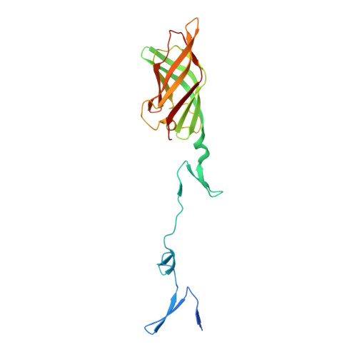
The GM2 Glycan Serves as a Functional Coreceptor for Serotype 1 Reovirus.

Reiss, K.Stencel, J.E.Liu, Y.Blaum, B.S.Reiter, D.M.Feizi, T.Dermody, T.S.Stehle, T.

(2012) PLoS Pathog 8: e1003078-e1003078

  • DOI: https://doi.org/10.1371/journal.ppat.1003078
  • Primary Citation of Related Structures:  
    4GU3, 4GU4

  • PubMed Abstract: 

    Viral attachment to target cells is the first step in infection and also serves as a determinant of tropism. Like many viruses, mammalian reoviruses bind with low affinity to cell-surface carbohydrate receptors to initiate the infectious process. Reoviruses disseminate with serotype-specific tropism in the host, which may be explained by differential glycan utilization. Although α2,3-linked sialylated oligosaccharides serve as carbohydrate receptors for type 3 reoviruses, neither a specific glycan bound by any reovirus serotype nor the function of glycan binding in type 1 reovirus infection was known. We have identified the oligosaccharide portion of ganglioside GM2 (the GM2 glycan) as a receptor for the attachment protein σ1 of reovirus strain type 1 Lang (T1L) using glycan array screening. The interaction of T1L σ1 with GM2 in solution was confirmed using NMR spectroscopy. We established that GM2 glycan engagement is required for optimal infection of mouse embryonic fibroblasts (MEFs) by T1L. Preincubation with GM2 specifically inhibited type 1 but not type 3 reovirus infection of MEFs. To provide a structural basis for these observations, we defined the mode of receptor recognition by determining the crystal structure of T1L σ1 in complex with the GM2 glycan. GM2 binds in a shallow groove in the globular head domain of T1L σ1. Both terminal sugar moieties of the GM2 glycan, N-acetylneuraminic acid and N-acetylgalactosamine, form contacts with the protein, providing an explanation for the observed specificity for GM2. Viruses with mutations in the glycan-binding domain display diminished hemagglutination capacity, a property dependent on glycan binding, and reduced capacity to infect MEFs. Our results define a novel mode of virus-glycan engagement and provide a mechanistic explanation for the serotype-dependent differences in glycan utilization by reovirus.


  • Organizational Affiliation

    Interfaculty Institute of Biochemistry, University of Tübingen, Tübingen, Germany.


Macromolecules
Find similar proteins by:  (by identity cutoff)  |  3D Structure
Entity ID: 1
MoleculeChains Sequence LengthOrganismDetailsImage
Outer capsid protein sigma-1
A, B, C
219Mammalian orthoreovirus 1 LangMutation(s): 0 
Gene Names: S1
UniProt
Find proteins for P04506 (Reovirus type 1 (strain Lang))
Explore P04506 
Go to UniProtKB:  P04506
Entity Groups  
Sequence Clusters30% Identity50% Identity70% Identity90% Identity95% Identity100% Identity
UniProt GroupP04506
Sequence Annotations
Expand
  • Reference Sequence
Oligosaccharides

Help

Entity ID: 2
MoleculeChains Length2D Diagram Glycosylation3D Interactions
N-acetyl-alpha-neuraminic acid-(2-3)-beta-D-galactopyranose-(1-4)-beta-D-glucopyranose
D, E
3N/A
Glycosylation Resources
GlyTouCan:  G91237TK
GlyCosmos:  G91237TK
GlyGen:  G91237TK
Entity ID: 3
MoleculeChains Length2D Diagram Glycosylation3D Interactions
N-acetyl-alpha-neuraminic acid-(2-3)-beta-D-galactopyranose
F
2N/A
Glycosylation Resources
GlyTouCan:  G30207PZ
GlyCosmos:  G30207PZ
GlyGen:  G30207PZ
Experimental Data & Validation

Experimental Data

  • Method: X-RAY DIFFRACTION
  • Resolution: 3.50 Å
  • R-Value Free: 0.197 
  • R-Value Work: 0.186 
  • R-Value Observed: 0.187 
  • Space Group: P 32 2 1
Unit Cell:
Length ( Å )Angle ( ˚ )
a = 149.43α = 90
b = 149.43β = 90
c = 165.19γ = 120
Software Package:
Software NamePurpose
XDSdata scaling
PHASERphasing
BUSTERrefinement
XDSdata reduction
XSCALEdata scaling

Structure Validation

View Full Validation Report


Entry History 

Deposition Data

Revision History  (Full details and data files)

  • Version 1.0: 2012-12-05
    Type: Initial release
  • Version 1.1: 2012-12-26
    Changes: Database references
  • Version 2.0: 2020-07-29
    Type: Remediation
    Reason: Carbohydrate remediation
    Changes: Atomic model, Data collection, Database references, Derived calculations, Structure summary
  • Version 2.1: 2024-02-28
    Changes: Data collection, Database references, Structure summary