


Crystal structures of the native, substrate- bound and inhibited forms of Mycobacterium tuberculosis imidazole glycerol phosphate dehydratase
Ahangar, M.S., Vyas, R., Nasir, N., Biswal, B.K.(2013) Acta Crystallogr D Biol Crystallogr
Experimental Data Snapshot
wwPDB Validation 3D Report Full Report

(2013) Acta Crystallogr D Biol Crystallogr
Entity ID: 1 | |||||
|---|---|---|---|---|---|
| Molecule | Chains | Sequence Length | Organism | Details | Image |
| Imidazoleglycerol-phosphate dehydratase | 216 | Mycobacterium tuberculosis | Mutation(s): 0 Gene Names: hisB, MT1637, MTCY336.03c, Rv1601 EC: 4.2.1.19 | 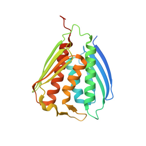 | |
UniProt | |||||
Find proteins for P9WML9 (Mycobacterium tuberculosis (strain ATCC 25618 / H37Rv)) Explore P9WML9 Go to UniProtKB: P9WML9 | |||||
Entity Groups | |||||
| Sequence Clusters | 30% Identity50% Identity70% Identity90% Identity95% Identity100% Identity | ||||
| UniProt Group | P9WML9 | ||||
Sequence AnnotationsExpand | |||||
| |||||
| Ligands 2 Unique | |||||
|---|---|---|---|---|---|
| ID | Chains | Name / Formula / InChI Key | 2D Diagram | 3D Interactions | |
| EDO Query on EDO | F [auth A] | 1,2-ETHANEDIOL C2 H6 O2 LYCAIKOWRPUZTN-UHFFFAOYSA-N |  | ||
| MN Query on MN | B [auth A], C [auth A], D [auth A], E [auth A] | MANGANESE (II) ION Mn WAEMQWOKJMHJLA-UHFFFAOYSA-N |  | ||
| Length ( Å ) | Angle ( ˚ ) |
|---|---|
| a = 112.539 | α = 90 |
| b = 112.539 | β = 90 |
| c = 112.539 | γ = 90 |
| Software Name | Purpose |
|---|---|
| StructureStudio | data collection |
| PHASER | phasing |
| REFMAC | refinement |
| HKL-2000 | data reduction |
| HKL-2000 | data scaling |
RCSB PDB (citation) is hosted by
RCSB PDB is a member of the

