4GNK

Crystal structure of Galphaq in complex with full-length human PLCbeta3


Experimental Data Snapshot

  • Method: X-RAY DIFFRACTION
  • Resolution: 4.00 Å
  • R-Value Free: 0.255 
  • R-Value Work: 0.214 
  • R-Value Observed: 0.216 

wwPDB Validation   3D Report Full Report


Ligand Structure Quality Assessment 


This is version 1.2 of the entry. See complete history


Literature

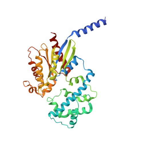
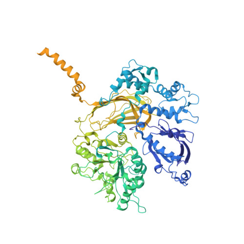
Full-length G alpha (q)-phospholipase C-beta 3 structure reveals interfaces of the C-terminal coiled-coil domain.

Lyon, A.M.Dutta, S.Boguth, C.A.Skiniotis, G.Tesmer, J.J.

(2013) Nat Struct Mol Biol 20: 355-362

  • DOI: https://doi.org/10.1038/nsmb.2497
  • Primary Citation of Related Structures:  
    4GNK

  • PubMed Abstract: 

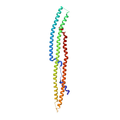
    Phospholipase C-β (PLCβ) is directly activated by Gαq, but the molecular basis for how its distal C-terminal domain (CTD) contributes to maximal activity is poorly understood. Herein we present both the crystal structure and cryo-EM three-dimensional reconstructions of human full-length PLCβ3 in complex with mouse Gαq. The distal CTD forms an extended monomeric helical bundle consisting of three antiparallel segments with structural similarity to membrane-binding bin-amphiphysin-Rvs (BAR) domains. Sequence conservation of the distal CTD suggests putative membrane and protein interaction sites, the latter of which bind the N-terminal helix of Gαq in both the crystal structure and cryo-EM reconstructions. Functional analysis suggests that the distal CTD has roles in membrane targeting and in optimizing the orientation of the catalytic core at the membrane for maximal rates of lipid hydrolysis.


  • Organizational Affiliation

    Life Sciences Institute, University of Michigan, Ann Arbor, Michigan, USA.


Macromolecules
Find similar proteins by:  (by identity cutoff)  |  3D Structure
Entity ID: 1
MoleculeChains Sequence LengthOrganismDetailsImage
Guanine nucleotide-binding protein G(q) subunit alpha
A, C
353Mus musculusMutation(s): 0 
Gene Names: g alpha qGnaq
Membrane Entity: Yes 
UniProt
Find proteins for P21279 (Mus musculus)
Explore P21279 
Go to UniProtKB:  P21279
Entity Groups  
Sequence Clusters30% Identity50% Identity70% Identity90% Identity95% Identity100% Identity
UniProt GroupP21279
Sequence Annotations
Expand
  • Reference Sequence
Find similar proteins by:  (by identity cutoff)  |  3D Structure
Entity ID: 2
MoleculeChains Sequence LengthOrganismDetailsImage
1-phosphatidylinositol 4,5-bisphosphate phosphodiesterase beta-3
B, D
1,235Homo sapiensMutation(s): 0 
Gene Names: phospholipase c beta 3PLCB3
EC: 3.1.4.11
Membrane Entity: Yes 
UniProt & NIH Common Fund Data Resources
Find proteins for Q01970 (Homo sapiens)
Explore Q01970 
Go to UniProtKB:  Q01970
PHAROS:  Q01970
GTEx:  ENSG00000149782 
Entity Groups  
Sequence Clusters30% Identity50% Identity70% Identity90% Identity95% Identity100% Identity
UniProt GroupQ01970
Sequence Annotations
Expand
  • Reference Sequence
Find similar proteins by:  (by identity cutoff)  |  3D Structure
Entity ID: 3
MoleculeChains Sequence LengthOrganismDetailsImage
1-phosphatidylinositol 4,5-bisphosphate phosphodiesterase beta-3259Homo sapiensMutation(s): 0 
Gene Names: phospholipase c beta 3PLCB3
Membrane Entity: Yes 
UniProt & NIH Common Fund Data Resources
Find proteins for Q01970 (Homo sapiens)
Explore Q01970 
Go to UniProtKB:  Q01970
PHAROS:  Q01970
GTEx:  ENSG00000149782 
Entity Groups  
Sequence Clusters30% Identity50% Identity70% Identity90% Identity95% Identity100% Identity
UniProt GroupQ01970
Sequence Annotations
Expand
  • Reference Sequence
Small Molecules
Ligands 4 Unique
IDChains Name / Formula / InChI Key2D Diagram3D Interactions
GDP
Query on GDP

Download Ideal Coordinates CCD File 
F [auth A],
J [auth C]
GUANOSINE-5'-DIPHOSPHATE
C10 H15 N5 O11 P2
QGWNDRXFNXRZMB-UUOKFMHZSA-N
ALF
Query on ALF

Download Ideal Coordinates CCD File 
G [auth A],
K [auth C]
TETRAFLUOROALUMINATE ION
Al F4
UYOMQIYKOOHAMK-UHFFFAOYSA-J
CA
Query on CA

Download Ideal Coordinates CCD File 
I [auth B],
M [auth D]
CALCIUM ION
Ca
BHPQYMZQTOCNFJ-UHFFFAOYSA-N
MG
Query on MG

Download Ideal Coordinates CCD File 
H [auth A],
L [auth C]
MAGNESIUM ION
Mg
JLVVSXFLKOJNIY-UHFFFAOYSA-N
Experimental Data & Validation

Experimental Data

  • Method: X-RAY DIFFRACTION
  • Resolution: 4.00 Å
  • R-Value Free: 0.255 
  • R-Value Work: 0.214 
  • R-Value Observed: 0.216 
  • Space Group: P 21 21 21
Unit Cell:
Length ( Å )Angle ( ˚ )
a = 91.423α = 90
b = 188.845β = 90
c = 293.857γ = 90
Software Package:
Software NamePurpose
HKL-2000data collection
PHASERphasing
REFMACrefinement
DENZOdata reduction
SCALEPACKdata scaling

Structure Validation

View Full Validation Report


Ligand Structure Quality Assessment 


Entry History 

Deposition Data

Revision History  (Full details and data files)

  • Version 1.0: 2013-02-06
    Type: Initial release
  • Version 1.1: 2013-09-04
    Changes: Database references
  • Version 1.2: 2023-09-13
    Changes: Data collection, Database references, Derived calculations, Refinement description