


The crystal structure of thymidylate kinase from Pseudomonas aeruginosa PAO1 in complex with AZT Monophosphate
Tan, K., Joachimiak, G., Jedrzejczak, R., Sacchettini, J., Joachimiak, A.To be published.
Experimental Data Snapshot
Entity ID: 1 | |||||
|---|---|---|---|---|---|
| Molecule | Chains | Sequence Length | Organism | Details | Image |
| Thymidylate kinase | 213 | Pseudomonas aeruginosa PAO1 | Mutation(s): 0 Gene Names: PA2962, tmk EC: 2.7.4.9 | 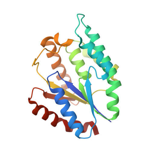 | |
UniProt | |||||
Find proteins for Q9HZN8 (Pseudomonas aeruginosa (strain ATCC 15692 / DSM 22644 / CIP 104116 / JCM 14847 / LMG 12228 / 1C / PRS 101 / PAO1)) Explore Q9HZN8 Go to UniProtKB: Q9HZN8 | |||||
Entity Groups | |||||
| Sequence Clusters | 30% Identity50% Identity70% Identity90% Identity95% Identity100% Identity | ||||
| UniProt Group | Q9HZN8 | ||||
Sequence AnnotationsExpand | |||||
| |||||
| Ligands 4 Unique | |||||
|---|---|---|---|---|---|
| ID | Chains | Name / Formula / InChI Key | 2D Diagram | 3D Interactions | |
| ATM Query on ATM | E [auth A], H [auth B], L [auth C], O [auth D] | 3'-AZIDO-3'-DEOXYTHYMIDINE-5'-MONOPHOSPHATE C10 H14 N5 O7 P OIFWQOKDSPDILA-XLPZGREQSA-N |  | ||
| GOL Query on GOL | G [auth A], J [auth B], K [auth B] | GLYCEROL C3 H8 O3 PEDCQBHIVMGVHV-UHFFFAOYSA-N |  | ||
| CA Query on CA | P [auth D] | CALCIUM ION Ca BHPQYMZQTOCNFJ-UHFFFAOYSA-N |  | ||
| CL Query on CL | F [auth A] I [auth B] M [auth C] N [auth C] Q [auth D] | CHLORIDE ION Cl VEXZGXHMUGYJMC-UHFFFAOYSA-M |  | ||
| Length ( Å ) | Angle ( ˚ ) |
|---|---|
| a = 44.5 | α = 90 |
| b = 123.404 | β = 100 |
| c = 73.92 | γ = 90 |
| Software Name | Purpose |
|---|---|
| SBC-Collect | data collection |
| MOLREP | phasing |
| PHENIX | refinement |
| HKL-3000 | data reduction |
| HKL-3000 | data scaling |
RCSB PDB (citation) is hosted by
RCSB PDB is a member of the

