4GI3

Crystal structure of Greglin in complex with subtilisin


Experimental Data Snapshot

  • Method: X-RAY DIFFRACTION
  • Resolution: 1.75 Å
  • R-Value Free: 0.201 
  • R-Value Work: 0.166 
  • R-Value Observed: 0.168 

wwPDB Validation   3D Report Full Report


This is version 1.2 of the entry. See complete history


Literature

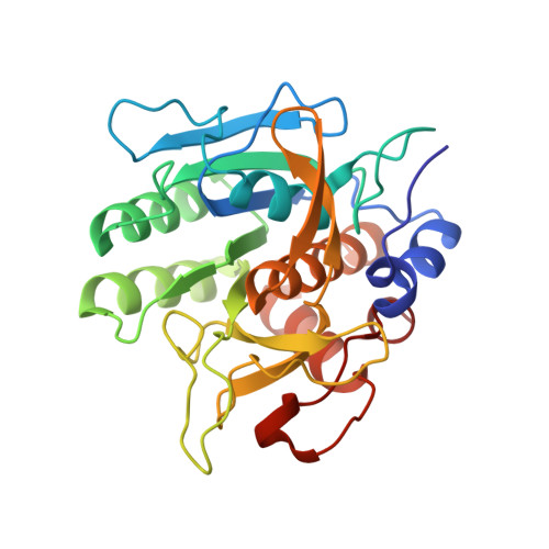
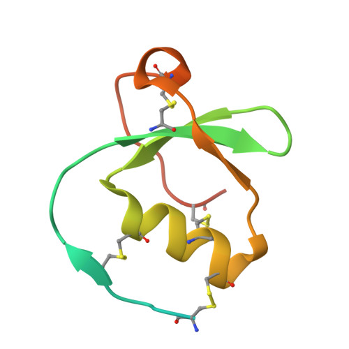
Crystal structure of greglin, a novel non-classical Kazal inhibitor, in complex with subtilisin

Derache, C.Epinette, C.Roussel, A.Gabant, G.Cadene, M.Korkmaz, B.Gauthier, F.Kellenberger, C.

(2012) FEBS J 279: 4466-4478

  • DOI: https://doi.org/10.1111/febs.12033
  • Primary Citation of Related Structures:  
    4GI3

  • PubMed Abstract: 

    Greglin is an 83-residue serine protease inhibitor purified from the ovaries of the locust Schistocerca gregaria. Greglin is a strong inhibitor of subtilisin and human neutrophil elastase, acting at sub-nanomolar and nanomolar concentrations, respectively; it also inhibits neutrophil cathepsin G, α-chymotrypsin and porcine pancreatic elastase, but to a lesser extent. In the present study, we show that greglin resists denaturation at high temperature (95 °C) and after exposure to acetonitrile and acidic or basic pH. Greglin is composed of two domains consisting of residues 1-20 and 21-83. Mass spectrometry indicates that the N-terminal domain (1-20) is post-translationally modified by phosphorylations at three sites and probably contains a glycosylation site. The crystal structure of the region of greglin comprising residues 21-78 in complex with subtilisin was determined at 1.75 Å resolution. Greglin represents a novel member of the non-classical Kazal inhibitors, as it has a unique additional C-terminal region (70-83) connected to the core of the molecule via a supplementary disulfide bond. The stability of greglin was compared with that of an ovomucoid inhibitor. The thermostability and inhibitory specificity of greglin are discussed in light of its structure. In particular, we propose that the C-terminal region is responsible for non-favourable interactions with the autolysis loop (140-loop) of serine proteases of the chymotrypsin family, and thus governs specificity.


  • Organizational Affiliation

    Centre de Biophysique Moléculaire, UPR 4301 CNRS conventionnée avec l'Université d'Orléans, Orléans Cedex 2, France.


Macromolecules
Find similar proteins by:  (by identity cutoff)  |  3D Structure
Entity ID: 1
MoleculeChains Sequence LengthOrganismDetailsImage
KerA275Bacillus licheniformisMutation(s): 0 
UniProt
Find proteins for Q9FDF2 (Bacillus licheniformis)
Explore Q9FDF2 
Go to UniProtKB:  Q9FDF2
Entity Groups  
Sequence Clusters30% Identity50% Identity70% Identity90% Identity95% Identity100% Identity
UniProt GroupQ9FDF2
Sequence Annotations
Expand
  • Reference Sequence
Find similar proteins by:  (by identity cutoff)  |  3D Structure
Entity ID: 2
MoleculeChains Sequence LengthOrganismDetailsImage
GreglinB [auth C]83Schistocerca gregariaMutation(s): 0 
UniProt
Find proteins for P85064 (Schistocerca gregaria)
Explore P85064 
Go to UniProtKB:  P85064
Entity Groups  
Sequence Clusters30% Identity50% Identity70% Identity90% Identity95% Identity100% Identity
UniProt GroupP85064
Sequence Annotations
Expand
  • Reference Sequence
Experimental Data & Validation

Experimental Data

  • Method: X-RAY DIFFRACTION
  • Resolution: 1.75 Å
  • R-Value Free: 0.201 
  • R-Value Work: 0.166 
  • R-Value Observed: 0.168 
  • Space Group: C 1 2 1
Unit Cell:
Length ( Å )Angle ( ˚ )
a = 131.07α = 90
b = 39.708β = 98.05
c = 59.75γ = 90
Software Package:
Software NamePurpose
DNAdata collection
AMoREphasing
BUSTERrefinement
MOSFLMdata reduction
SCALAdata scaling

Structure Validation

View Full Validation Report


Entry History 

Deposition Data

Revision History  (Full details and data files)

  • Version 1.0: 2012-11-14
    Type: Initial release
  • Version 1.1: 2013-09-04
    Changes: Database references
  • Version 1.2: 2023-11-08
    Changes: Data collection, Database references, Refinement description