4GCP

Crystal Structure of E. coli OmpF porin in complex with Ampicillin


Experimental Data Snapshot

  • Method: X-RAY DIFFRACTION
  • Resolution: 1.98 Å
  • R-Value Free: 0.239 
  • R-Value Work: 0.203 
  • R-Value Observed: 0.205 

wwPDB Validation   3D Report Full Report


Ligand Structure Quality Assessment 


This is version 1.2 of the entry. See complete history


Literature

The Binding of Antibiotics in OmpF Porin.

Ziervogel, B.K.Roux, B.

(2013) Structure 21: 76-87

  • DOI: https://doi.org/10.1016/j.str.2012.10.014
  • Primary Citation of Related Structures:  
    4GCP, 4GCQ, 4GCS

  • PubMed Abstract: 

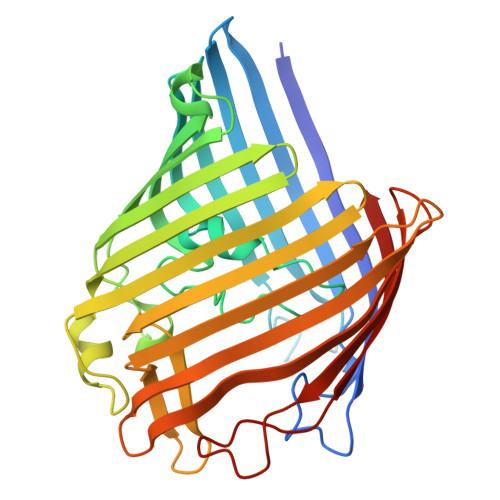
    The structure of OmpF porin in complex with three common antibiotics (zwitterionic ampicillin, anionic ertapenem, and di-anionic carbenicillin) was determined using X-ray crystallography. The three antibiotics are found to bind within the extracellular and periplasmic pore vestibules, away from the narrow OmpF constriction zone. Using the X-ray structures as a starting point, nonequilibrium molecular dynamics simulations with an applied membrane voltage show that ionic current through the OmpF channel is blocked with bound ampicillin, but not with bound carbenicillin. The susceptibility of Escherichia coli expressing OmpF mutants to ampicillin and carbenicillin was also experimentally characterized using microbiologic assays. These results show that general diffusion by OmpF porins allows for transfer of molecules with varied charged states and give insights into the design of more efficient antibiotics. A better understanding of this mechanism will shed light on nature's way of devising channels able to enhance the transport of molecules through membranes.


  • Organizational Affiliation

    Department of Biochemistry and Molecular Biology, Gordon Center for Integrative Science, The University of Chicago, 929 East 57(th) Street, Chicago, IL 60637, USA.


Macromolecules
Find similar proteins by:  (by identity cutoff)  |  3D Structure
Entity ID: 1
MoleculeChains Sequence LengthOrganismDetailsImage
Outer membrane protein F
A, B
341Escherichia coli K-12Mutation(s): 0 
Gene Names: ompFcmlBcoacrytolFb0929JW0912
Membrane Entity: Yes 
UniProt
Find proteins for P02931 (Escherichia coli (strain K12))
Explore P02931 
Go to UniProtKB:  P02931
Entity Groups  
Sequence Clusters30% Identity50% Identity70% Identity90% Identity95% Identity100% Identity
UniProt GroupP02931
Sequence Annotations
Expand
  • Reference Sequence
Small Molecules
Ligands 1 Unique
IDChains Name / Formula / InChI Key2D Diagram3D Interactions
AIC
Query on AIC

Download Ideal Coordinates CCD File 
C [auth A],
D [auth B]
(2S,5R,6R)-6-{[(2R)-2-AMINO-2-PHENYLETHANOYL]AMINO}-3,3-DIMETHYL-7-OXO-4-THIA-1-AZABICYCLO[3.2.0]HEPTANE-2-CARBOXYLIC ACID
C16 H19 N3 O4 S
AVKUERGKIZMTKX-NJBDSQKTSA-N
Experimental Data & Validation

Experimental Data

  • Method: X-RAY DIFFRACTION
  • Resolution: 1.98 Å
  • R-Value Free: 0.239 
  • R-Value Work: 0.203 
  • R-Value Observed: 0.205 
  • Space Group: P 3
Unit Cell:
Length ( Å )Angle ( ˚ )
a = 116.6α = 90
b = 116.6β = 90
c = 53.27γ = 120
Software Package:
Software NamePurpose
HKL-2000data collection
PHASERphasing
REFMACrefinement
MOSFLMdata reduction
SCALAdata scaling

Structure Validation

View Full Validation Report


Ligand Structure Quality Assessment 


Entry History 

Deposition Data

Revision History  (Full details and data files)

  • Version 1.0: 2012-12-19
    Type: Initial release
  • Version 1.1: 2013-02-06
    Changes: Database references
  • Version 1.2: 2024-02-28
    Changes: Data collection, Database references, Derived calculations, Refinement description