4GAW

Crystal structure of active human granzyme H


Experimental Data Snapshot

  • Method: X-RAY DIFFRACTION
  • Resolution: 3.00 Å
  • R-Value Free: 0.306 
  • R-Value Work: 0.271 
  • R-Value Observed: 0.272 

wwPDB Validation   3D Report Full Report


This is version 1.2 of the entry. See complete history


Literature

Identification of SERPINB1 as a physiological inhibitor of human granzyme H

Wang, L.Li, Q.Wu, L.Liu, S.Zhang, Y.Yang, X.Zhu, P.Zhang, H.Zhang, K.Lou, J.Liu, P.Tong, L.Sun, F.Fan, Z.

(2013) J Immunol 190: 1319-1330

  • DOI: https://doi.org/10.4049/jimmunol.1202542
  • Primary Citation of Related Structures:  
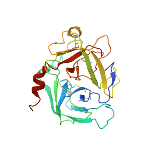
    4GA7, 4GAW

  • PubMed Abstract: 

    The granzyme/perforin pathway is a major mechanism for cytotoxic lymphocytes to eliminate virus-infected and tumor cells. The balance between activation and inhibition of the proteolytic cascade must be tightly controlled to avoid self damage. Granzyme H (GzmH) is constitutively expressed in NK cells and induces target cell death; however, how GzmH activity is regulated remains elusive. We reported earlier the crystal structures of inactive D102N-GzmH alone and in complex with its synthetic substrate and inhibitor, as well as defined the mechanisms of substrate recognition and enzymatic activation. In this study, we identified SERPINB1 as a potent intracellular inhibitor for GzmH. Upon cleavage of the reactive center loop at Phe(343), SERPINB1 forms an SDS-stable covalent complex with GzmH. SERPINB1 overexpression suppresses GzmH- or LAK cell-mediated cytotoxicity. We determined the crystal structures of active GzmH and SERPINB1 (LM-DD mutant) in the native conformation to 3.0- and 2.9-Å resolution, respectively. Molecular modeling reveals the possible conformational changes in GzmH for the suicide inhibition. Our findings provide new insights into the inhibitory mechanism of SERPINB1 against human GzmH.


  • Organizational Affiliation

    Chinese Academy of Sciences Key Laboratory of Infection and Immunity, Institute of Biophysics, Chinese Academy of Sciences, Beijing 100101, China.


Macromolecules
Find similar proteins by:  (by identity cutoff)  |  3D Structure
Entity ID: 1
MoleculeChains Sequence LengthOrganismDetailsImage
Granzyme H
A, B, C, D, E
A, B, C, D, E, F, G, H, I, J, K, L
226Homo sapiensMutation(s): 0 
Gene Names: GZMHCGL2CTSGL2
EC: 3.4.21
UniProt & NIH Common Fund Data Resources
Find proteins for P20718 (Homo sapiens)
Explore P20718 
Go to UniProtKB:  P20718
PHAROS:  P20718
GTEx:  ENSG00000100450 
Entity Groups  
Sequence Clusters30% Identity50% Identity70% Identity90% Identity95% Identity100% Identity
UniProt GroupP20718
Sequence Annotations
Expand
  • Reference Sequence
Small Molecules
Ligands 2 Unique
IDChains Name / Formula / InChI Key2D Diagram3D Interactions
SO4
Query on SO4

Download Ideal Coordinates CCD File 
AA [auth I]
CA [auth L]
DA [auth L]
M [auth B]
N [auth B]
AA [auth I],
CA [auth L],
DA [auth L],
M [auth B],
N [auth B],
O [auth B],
P [auth B],
R [auth D],
S [auth D],
T [auth D],
V [auth E],
W [auth F],
X [auth F],
Y [auth F],
Z [auth I]
SULFATE ION
O4 S
QAOWNCQODCNURD-UHFFFAOYSA-L
CL
Query on CL

Download Ideal Coordinates CCD File 
BA [auth I],
Q [auth C],
U [auth D]
CHLORIDE ION
Cl
VEXZGXHMUGYJMC-UHFFFAOYSA-M
Experimental Data & Validation

Experimental Data

  • Method: X-RAY DIFFRACTION
  • Resolution: 3.00 Å
  • R-Value Free: 0.306 
  • R-Value Work: 0.271 
  • R-Value Observed: 0.272 
  • Space Group: P 21 21 2
Unit Cell:
Length ( Å )Angle ( ˚ )
a = 170.369α = 90
b = 367.07β = 90
c = 61.137γ = 90
Software Package:
Software NamePurpose
ADSCdata collection
COMOphasing
REFMACrefinement
HKL-2000data reduction
HKL-2000data scaling

Structure Validation

View Full Validation Report


Entry History 

Deposition Data

Revision History  (Full details and data files)

  • Version 1.0: 2013-01-16
    Type: Initial release
  • Version 1.1: 2013-07-17
    Changes: Database references
  • Version 1.2: 2023-11-08
    Changes: Data collection, Database references, Derived calculations, Refinement description