Structure analysis of archaeal AMP phosphorylase reveals two unique modes of dimerization
Nishitani, Y., Aono, R., Nakamura, A., Sato, T., Atomi, H., Imanaka, T., Miki, K.(2013) J Mol Biol 425: 2709-2721
- PubMed: 23659790
- DOI: https://doi.org/10.1016/j.jmb.2013.04.026
- Primary Citation of Related Structures:
4GA4, 4GA5, 4GA6 - PubMed Abstract:
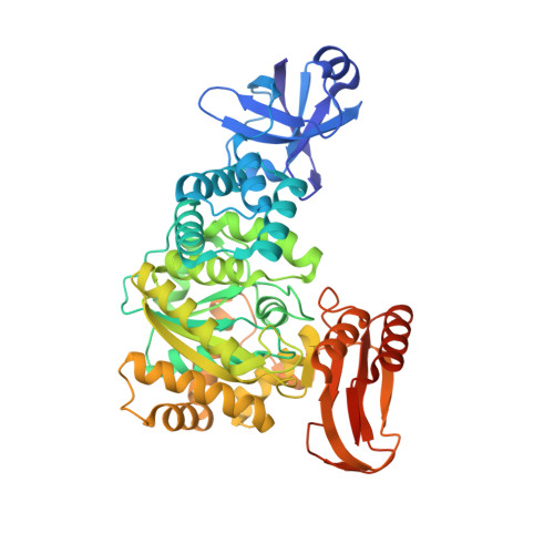
AMP phosphorylase (AMPpase) catalyzes the initial reaction in a novel AMP metabolic pathway recently found in archaea, converting AMP and phosphate into adenine and ribose 1,5-bisphosphate. Gel-filtration chromatography revealed that AMPpase from Thermococcus kodakarensis (Tk-AMPpase) forms an exceptionally large macromolecular structure (>40-mers) in solution. To investigate its unique multimerization feature, we determined the first crystal structures of Tk-AMPpase, in the apo-form and in complex with substrates. Structures of two truncated forms of Tk-AMPpase (Tk-AMPpaseΔN84 and Tk-AMPpaseΔC10) clarified that this multimerization is achieved by two dimer interfaces within a single molecule: one by the central domain and the other by the C-terminal domain, which consists of an unexpected domain-swapping interaction. The N-terminal domain, characteristic of archaeal enzymes, is essential for enzymatic activity, participating in multimerization as well as domain closure of the active site upon substrate binding. Moreover, biochemical analysis demonstrated that the macromolecular assembly of Tk-AMPpase contributes to its high thermostability, essential for an enzyme from a hyperthermophile. Our findings unveil a unique archaeal nucleotide phosphorylase that is distinct in both function and structure from previously known members of the nucleoside phosphorylase II family.
Organizational Affiliation:
Department of Chemistry, Graduate School of Science, Kyoto University, Sakyo-ku, Kyoto 606-8502, Japan.