4G68

Biochemical and structural insights into xylan utilization by the thermophilic bacteriumcaldanaerobius polysaccharolyticus


Experimental Data Snapshot

  • Method: X-RAY DIFFRACTION
  • Resolution: 1.80 Å
  • R-Value Free: 0.261 
  • R-Value Work: 0.230 
  • R-Value Observed: 0.231 

wwPDB Validation   3D Report Full Report


This is version 2.0 of the entry. See complete history


Literature

Biochemical and Structural Insights into Xylan Utilization by the Thermophilic Bacterium Caldanaerobius polysaccharolyticus.

Han, Y.Agarwal, V.Dodd, D.Kim, J.Bae, B.Mackie, R.I.Nair, S.K.Cann, I.K.

(2012) J Biol Chem 287: 34946-34960

  • DOI: https://doi.org/10.1074/jbc.M112.391532
  • Primary Citation of Related Structures:  
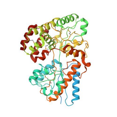
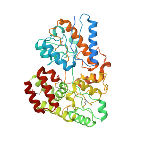
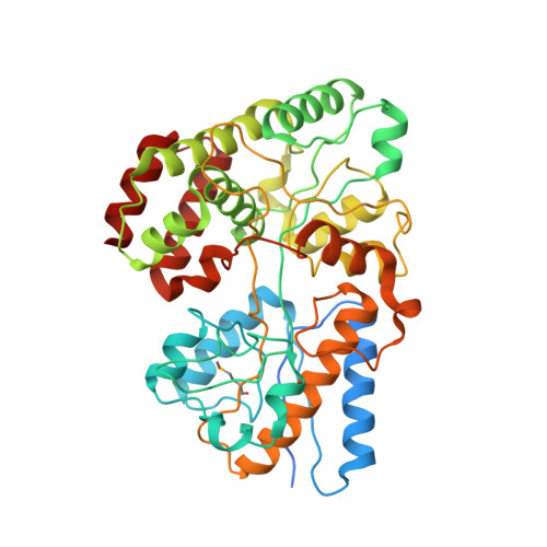
    4G68

  • PubMed Abstract: 

    Hemicellulose is the next most abundant plant cell wall component after cellulose. The abundance of hemicellulose such as xylan suggests that their hydrolysis and conversion to biofuels can improve the economics of bioenergy production. In an effort to understand xylan hydrolysis at high temperatures, we sequenced the genome of the thermophilic bacterium Caldanaerobius polysaccharolyticus. Analysis of the partial genome sequence revealed a gene cluster that contained both hydrolytic enzymes and also enzymes key to the pentose-phosphate pathway. The hydrolytic enzymes in the gene cluster were demonstrated to convert products from a large endoxylanase (Xyn10A) predicted to anchor to the surface of the bacterium. We further use structural and calorimetric studies to demonstrate that the end products of Xyn10A hydrolysis of xylan are recognized and bound by XBP1, a putative solute-binding protein, likely for transport into the cell. The XBP1 protein showed preference for xylo-oligosaccharides as follows: xylotriose > xylobiose > xylotetraose. To elucidate the structural basis for the oligosaccharide preference, we solved the co-crystal structure of XBP1 complexed with xylotriose to a 1.8-Å resolution. Analysis of the biochemical data in the context of the co-crystal structure reveals the molecular underpinnings of oligosaccharide length specificity.


  • Organizational Affiliation

    Energy Biosciences Institute, University of Illinois, Urbana, Illinois 61801; Institute for Genomic Biology, University of Illinois, Urbana, Illinois 61801.


Macromolecules
Find similar proteins by:  (by identity cutoff)  |  3D Structure
Entity ID: 1
MoleculeChains Sequence LengthOrganismDetailsImage
ABC transporter456CaldanaerobiusMutation(s): 0 
UniProt
Find proteins for J9PBT4 (Caldanaerobius)
Explore J9PBT4 
Go to UniProtKB:  J9PBT4
Entity Groups  
Sequence Clusters30% Identity50% Identity70% Identity90% Identity95% Identity100% Identity
UniProt GroupJ9PBT4
Sequence Annotations
Expand
  • Reference Sequence
Find similar proteins by:  (by identity cutoff)  |  3D Structure
Entity ID: 2
MoleculeChains Sequence LengthOrganismDetailsImage
ABC transporter432CaldanaerobiusMutation(s): 0 
UniProt
Find proteins for J9PBT5 (Caldanaerobius)
Explore J9PBT5 
Go to UniProtKB:  J9PBT5
Entity Groups  
Sequence Clusters30% Identity50% Identity70% Identity90% Identity95% Identity100% Identity
UniProt GroupJ9PBT5
Sequence Annotations
Expand
  • Reference Sequence
Find similar proteins by:  (by identity cutoff)  |  3D Structure
Entity ID: 3
MoleculeChains Sequence LengthOrganismDetailsImage
ABC transporter432CaldanaerobiusMutation(s): 0 
UniProt
Find proteins for J9PBT5 (Caldanaerobius)
Explore J9PBT5 
Go to UniProtKB:  J9PBT5
Entity Groups  
Sequence Clusters30% Identity50% Identity70% Identity90% Identity95% Identity100% Identity
UniProt GroupJ9PBT5
Sequence Annotations
Expand
  • Reference Sequence
Oligosaccharides

Help

Entity ID: 4
MoleculeChains Length2D Diagram Glycosylation3D Interactions
beta-D-xylopyranose-(1-4)-beta-D-xylopyranose-(1-4)-alpha-D-xylopyranose
D, E
3N/A
Glycosylation Resources
GlyTouCan:  G13162YT
GlyCosmos:  G13162YT
Small Molecules
Modified Residues  1 Unique
IDChains TypeFormula2D DiagramParent
MSE
Query on MSE
A
L-PEPTIDE LINKINGC5 H11 N O2 SeMET
Binding Affinity Annotations 
IDSourceBinding Affinity
XYP PDBBind:  4G68 Kd: 12 (nM) from 1 assay(s)
Experimental Data & Validation

Experimental Data

  • Method: X-RAY DIFFRACTION
  • Resolution: 1.80 Å
  • R-Value Free: 0.261 
  • R-Value Work: 0.230 
  • R-Value Observed: 0.231 
  • Space Group: P 21 21 21
Unit Cell:
Length ( Å )Angle ( ˚ )
a = 59.33α = 90
b = 150.862β = 90
c = 150.879γ = 90
Software Package:
Software NamePurpose
DENZOdata reduction
SCALEPACKdata scaling
REFMACrefinement
PDB_EXTRACTdata extraction

Structure Validation

View Full Validation Report


Entry History 

Deposition Data

Revision History  (Full details and data files)

  • Version 1.0: 2012-08-22
    Type: Initial release
  • Version 1.1: 2017-11-15
    Changes: Refinement description
  • Version 2.0: 2020-07-29
    Type: Remediation
    Reason: Carbohydrate remediation
    Changes: Advisory, Atomic model, Data collection, Derived calculations, Structure summary