4G37

Structure of cross-linked firefly luciferase in second catalytic conformation


Experimental Data Snapshot

  • Method: X-RAY DIFFRACTION
  • Resolution: 2.40 Å
  • R-Value Free: 0.259 
  • R-Value Work: 0.199 
  • R-Value Observed: 0.202 

wwPDB Validation   3D Report Full Report


Ligand Structure Quality Assessment 


This is version 1.2 of the entry. See complete history


Literature

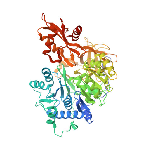
Crystal structure of firefly luciferase in a second catalytic conformation supports a domain alternation mechanism.

Sundlov, J.A.Fontaine, D.M.Southworth, T.L.Branchini, B.R.Gulick, A.M.

(2012) Biochemistry 51: 6493-6495

  • DOI: https://doi.org/10.1021/bi300934s
  • Primary Citation of Related Structures:  
    4G36, 4G37

  • PubMed Abstract: 

    Beetle luciferases catalyze a two-step reaction that includes the initial adenylation of the luciferin substrate, followed by an oxidative decarboxylation that ultimately produces light. Evidence for homologous acyl-CoA synthetases supports a domain alternation catalytic mechanism in which these enzymes' C-terminal domain rotates by ~140° to adopt two conformations that are used to catalyze the two partial reactions. While many structures exist of acyl-CoA synthetases in both conformations, to date only biochemical evidence supports domain alternation with luciferase. We have determined the structure of a cross-linked luciferase enzyme that is trapped in the second conformation. This new structure supports the role of the second catalytic conformation and provides insights into the biochemical mechanism of the luciferase oxidative step.


  • Organizational Affiliation

    Hauptman-Woodward Institute, 700 Ellicott Street, Buffalo, NY 14203, USA.


Macromolecules
Find similar proteins by:  (by identity cutoff)  |  3D Structure
Entity ID: 1
MoleculeChains Sequence LengthOrganismDetailsImage
Luciferin 4-monooxygenase
A, B
555Photinus pyralisMutation(s): 11 
EC: 1.13.12.7
UniProt
Find proteins for P08659 (Photinus pyralis)
Explore P08659 
Go to UniProtKB:  P08659
Entity Groups  
Sequence Clusters30% Identity50% Identity70% Identity90% Identity95% Identity100% Identity
UniProt GroupP08659
Sequence Annotations
Expand
  • Reference Sequence
Small Molecules
Ligands 4 Unique
IDChains Name / Formula / InChI Key2D Diagram3D Interactions
SLU
Query on SLU

Download Ideal Coordinates CCD File 
C [auth A],
G [auth B]
5'-O-[N-(DEHYDROLUCIFERYL)-SULFAMOYL] ADENOSINE
C21 H18 N8 O8 S3
LJLYTUYNVSHXQB-SOONXTGKSA-N
XLX
Query on XLX

Download Ideal Coordinates CCD File 
D [auth A],
H [auth B]
4,4'-(ethylenediimino)bis[4-oxobutyrate]
C11 H18 N2 O6
JUHMDCQKNGDTCV-UHFFFAOYSA-N
SO4
Query on SO4

Download Ideal Coordinates CCD File 
E [auth A],
F [auth A],
J [auth B],
K [auth B]
SULFATE ION
O4 S
QAOWNCQODCNURD-UHFFFAOYSA-L
CL
Query on CL

Download Ideal Coordinates CCD File 
I [auth B]CHLORIDE ION
Cl
VEXZGXHMUGYJMC-UHFFFAOYSA-M
Experimental Data & Validation

Experimental Data

  • Method: X-RAY DIFFRACTION
  • Resolution: 2.40 Å
  • R-Value Free: 0.259 
  • R-Value Work: 0.199 
  • R-Value Observed: 0.202 
  • Space Group: C 2 2 21
Unit Cell:
Length ( Å )Angle ( ˚ )
a = 75.638α = 90
b = 184.091β = 90
c = 170.53γ = 90
Software Package:
Software NamePurpose
Blu-Icedata collection
PHASERphasing
PHENIXrefinement
HKL-2000data reduction
SCALEPACKdata scaling

Structure Validation

View Full Validation Report


Ligand Structure Quality Assessment 


Entry History 

Deposition Data

Revision History  (Full details and data files)

  • Version 1.0: 2012-08-15
    Type: Initial release
  • Version 1.1: 2012-10-31
    Changes: Database references
  • Version 1.2: 2023-09-13
    Changes: Advisory, Data collection, Database references, Derived calculations, Refinement description